Karabulut Derya, Akin Ali Tugrul, Unsal Murat, Lekesizcan Ayça, Ozyazgan Tuğçe Merve, Keti Didem Barlak, Yakan Birkan, Ekebas Görkem
Department of Histology-Embryology, Faculty of Medicine, Erciyes University, Kayseri, Turkey.
Department of Biology, Faculty of Science, Erciyes University, Kayseri, Turkey.
Iran J Basic Med Sci. 2021 Feb;24(2):184-190. doi: 10.22038/IJBMS.2020.47711.10990.
Carbon tetrachloride (CCL) toxicity triggers fibrosis, activating various mechanisms within the cell. We aimed to create damage with CCL and investigate the effectiveness of L-carnitine on the mechanisms we identified.
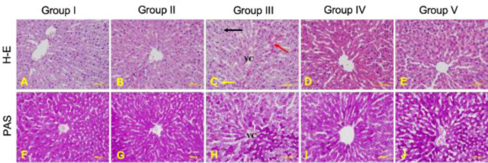
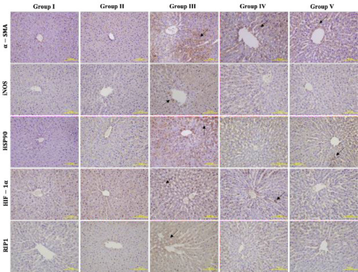
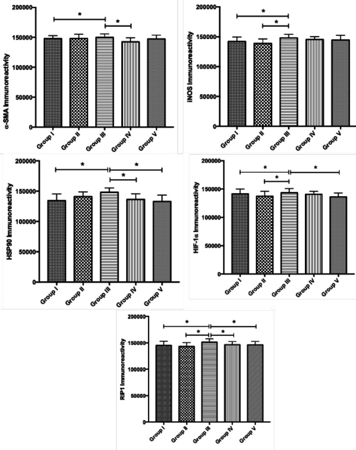
Forty rats were divided into 5 groups with equal number of rats in each group. Group I: Control group, Group II: L-carnitine group, 200 mg/kg L-carnitine twice a week, Group III: CCL group, 0.2 ml/100 gr CCL, IP, dissolved in olive oil 2 times a week during 6 weeks; Group IV: L-carnitine + CCL group, 200 mg/kg L-carnitine 24 hr before 0.2 ml/100 g CCL application twice a week; Group V: CCL + L-carnitine, 200 mg/kg L-carnitine half an hour after 0.2 ml/100 g CCL application. The liver was evaluated histologically. Immunohistochemically stained with α-SMA, iNOS, HSP90, HIF-1α, and RIP1. TNF-α, TGF-β, AST, ALT, ALP, and GGT measurements were evaluated.
In the classical lobule periphery, an increase in lipid accumulation and a decrease in glycogen accumulation were observed. After immunohistochemical measurements and biochemical analyzes, an increase in the expression density of all proteins was observed in group III. In group IV and V, an improvement in tissue and a decrease in protein expression densities were observed.
iNOS serves as a free radical scavenger in response to damage caused by increased toxicity of α-SMA, HSP90, and HIF-1α. Especially, increased RIP1 level in the tissue indicates the presence of necrosis in the tissue after CCL-toxicity. Supplementing the amount of endogenous L-carnitine with supplementation provides a significant improvement in the tissue.
四氯化碳(CCL)毒性引发纤维化,激活细胞内多种机制。我们旨在用CCL造成损伤并研究左旋肉碱对我们所确定机制的有效性。
40只大鼠分为5组,每组大鼠数量相等。第一组:对照组;第二组:左旋肉碱组,每周两次给予200mg/kg左旋肉碱;第三组:CCL组,腹腔注射0.2ml/100g CCL(溶于橄榄油),每周2次,共6周;第四组:左旋肉碱+CCL组,在每周两次给予0.2ml/100g CCL前24小时给予200mg/kg左旋肉碱;第五组:CCL+左旋肉碱组,在给予0.2ml/100g CCL后半小时给予200mg/kg左旋肉碱。对肝脏进行组织学评估。用α-SMA、诱导型一氧化氮合酶(iNOS)、热休克蛋白90(HSP90)、缺氧诱导因子-1α(HIF-1α)和受体相互作用蛋白1(RIP1)进行免疫组织化学染色。评估肿瘤坏死因子-α(TNF-α)、转化生长因子-β(TGF-β)、谷草转氨酶(AST)、谷丙转氨酶(ALT)、碱性磷酸酶(ALP)和γ-谷氨酰转肽酶(GGT)的测量值。
在经典肝小叶周边,观察到脂质蓄积增加和糖原蓄积减少。经过免疫组织化学测量和生化分析,第三组中所有蛋白质的表达密度均增加。在第四组和第五组中,观察到组织改善且蛋白质表达密度降低。
iNOS作为自由基清除剂,以应对由α-SMA、HSP90和HIF-1α毒性增加所导致的损伤。特别是,组织中RIP1水平升高表明CCL毒性后组织中存在坏死。补充内源性左旋肉碱的量可使组织有显著改善。