Department of Ophthalmology, Eye & ENT Hospital, Fudan University, Shanghai, China.
Shanghai Key Laboratory of Visual Impairment and Restoration, Fudan University, Shanghai, China.
Mol Vis. 2021 May 1;27:206-220. eCollection 2021.
To explore synaptic changes and the response of microglia in a light-induced photoreceptor degeneration model.
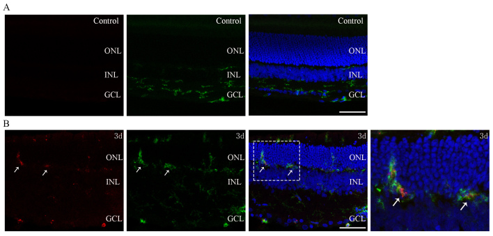
Sprague-Dawley rats were euthanized 1 h, 1 day, 3 days, 7 days, and 14 days after being exposed to intense blue light for 24 h. Hematoxylin and eosin (H&E) and terminal deoxynucleotidyl transferase dUTP nick-end labeling (TUNEL) staining were used to evaluate changes in the outer nuclear layer (ONL). Transmission electron microscopy (TEM) was applied to observe the ultrastructural changes in the synapses between the photoreceptors and second-order neurons. Western blotting was conducted to evaluate specific proteins, including postsynaptic density-95 (PSD-95), metabotropic glutamate receptor 6 (mGluR6), synapsin I, and synaptophysin. Immunofluorescence of CD11b and PKC-α or mGluR6 was used to explore the spatial relationships between microglial processes and synaptic elements. Immunoelectron microscopy of PSD-95 was performed to further confirm its engulfment of synaptic materials.
H&E and TUNEL staining showed that the thickness of the ONL decreased markedly, and the number of apoptotic photoreceptors peaked at day 1. TEM revealed darkened photoreceptor terminals and that ribbons of them were floating in the cytoplasm, coinciding with the downregulation of PSD-95 and mGluR6. Downstream synaptic protein synapsin I and synaptophysin exhibited upregulation in the inner plexiform layer. Activated microglia migrated to the outer retina, and their processes were found in close proximity to synapses in the outer plexiform layer under light and electron microscopy levels. Double immunostaining of CD11b and mGluR6 showed colocalization. PSD-95-immunoreactive electron-dense materials were observed inside the microglia suggesting engulfment of synaptic components.
The study showed that there are early synaptic impairment and late compensatory changes in downstream synapses in this photic injury model. Activated microglia touched and directly engulfed synaptic materials. Microglia may play a role or a partial role in synaptic changes.
探讨光诱导的光感受器变性模型中的突触变化和小胶质细胞的反应。
Sprague-Dawley 大鼠在暴露于强蓝光 24 小时后 1 小时、1 天、3 天、7 天和 14 天被安乐死。苏木精和伊红(H&E)和末端脱氧核苷酸转移酶 dUTP 缺口末端标记(TUNEL)染色用于评估外核层(ONL)的变化。透射电子显微镜(TEM)用于观察光感受器和二级神经元之间突触的超微结构变化。Western blot 用于评估包括突触后密度蛋白-95(PSD-95)、代谢型谷氨酸受体 6(mGluR6)、突触素 I 和突触小体蛋白在内的特定蛋白质。用 CD11b 和 PKC-α或 mGluR6 的免疫荧光来探索小胶质细胞突起与突触成分之间的空间关系。用 PSD-95 的免疫电镜进一步证实其吞噬突触物质。
H&E 和 TUNEL 染色显示,ONL 的厚度明显变薄,凋亡光感受器的数量在第 1 天达到峰值。TEM 显示光感受器末端变暗,它们的带状物漂浮在细胞质中,这与 PSD-95 和 mGluR6 的下调相一致。下游突触蛋白突触素 I 和突触小体蛋白在内丛状层中表达上调。活化的小胶质细胞迁移到外视网膜,在光镜和电镜水平下,其突起被发现与外丛状层中的突触紧密相邻。CD11b 和 mGluR6 的双重免疫染色显示共定位。在小胶质细胞内观察到 PSD-95 免疫反应性电子致密物质,提示吞噬突触成分。
该研究表明,在这种光损伤模型中存在早期突触损伤和晚期下游突触的代偿性变化。活化的小胶质细胞接触并直接吞噬突触物质。小胶质细胞可能在突触变化中发挥作用或部分作用。