Feng ChaoYi, Wang Xin, Liu TianJin, Zhang Meng, Xu GeZhi, Ni YingQin
Department of Ophthalmology, Eye & ENT Hospital of Fudan University, Shanghai, People's Republic of China.
Shanghai Key Laboratory of Visual Impairment and Restoration, Shanghai, People's Republic of China.
Mol Vis. 2017 Nov 1;23:765-777. eCollection 2017.
To explore the effect of the CCL2 and CCR2 system on the activation and migration of microglia and monocytes in light-induced photoreceptor apoptosis.
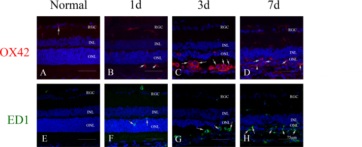
At 1 day, 3 days, 7 days, and 14 days after light exposure, OX42 and ED1 immunostaining were used to label the activation and migration of microglia and monocytes. Double immunostaining of CCL2 with terminal deoxynucleotidyl transferase dUTP nick end labeling (TUNEL), OX42, or glial fibrillary acidic protein (GFAP) was applied to explore the relationships among CCL2, apoptotic photoreceptors, activated microglia and monocytes, and macroglial cells (Müller cells and astrocytes). Real-time PCR was used to evaluate the mRNA levels of retinal CCL2 and CCR2 and the proinflammatory factors interleukin (IL)-1 beta and tumor necrosis factor (TNF)-alpha.
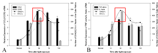
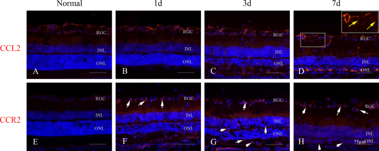
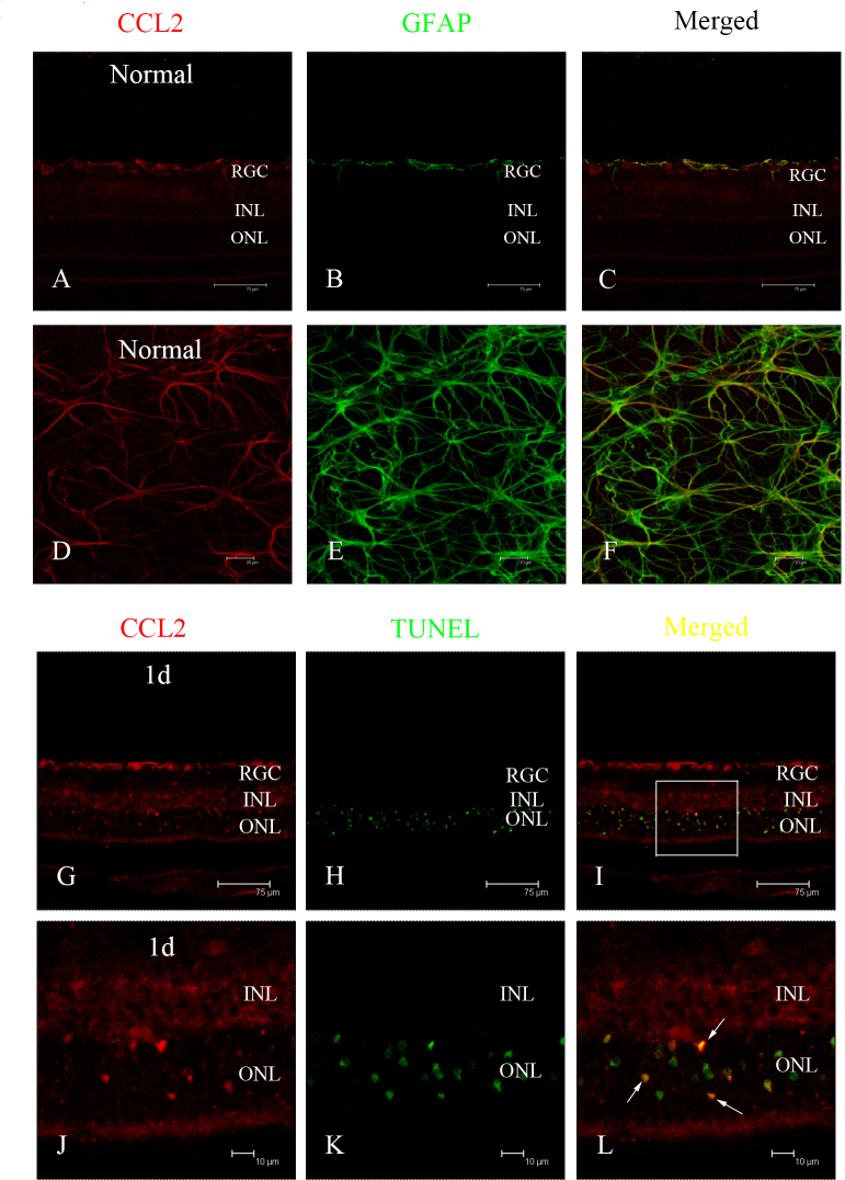
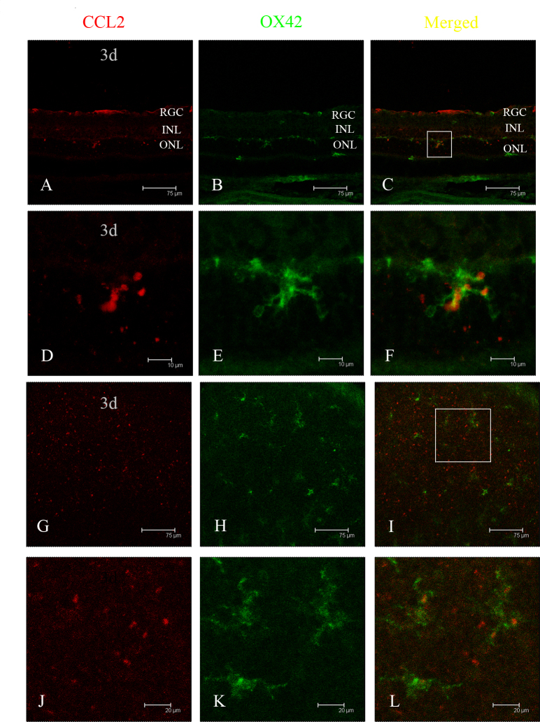
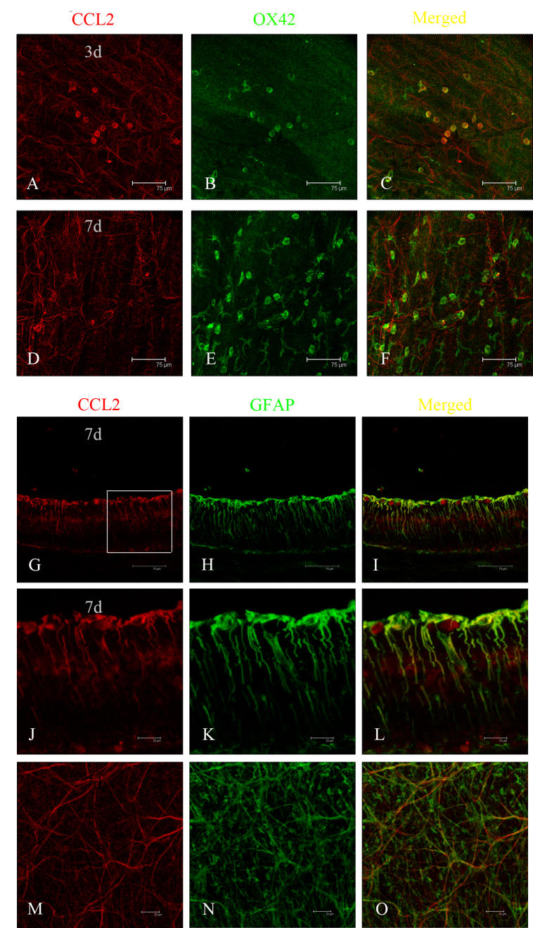
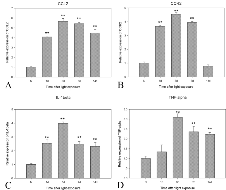
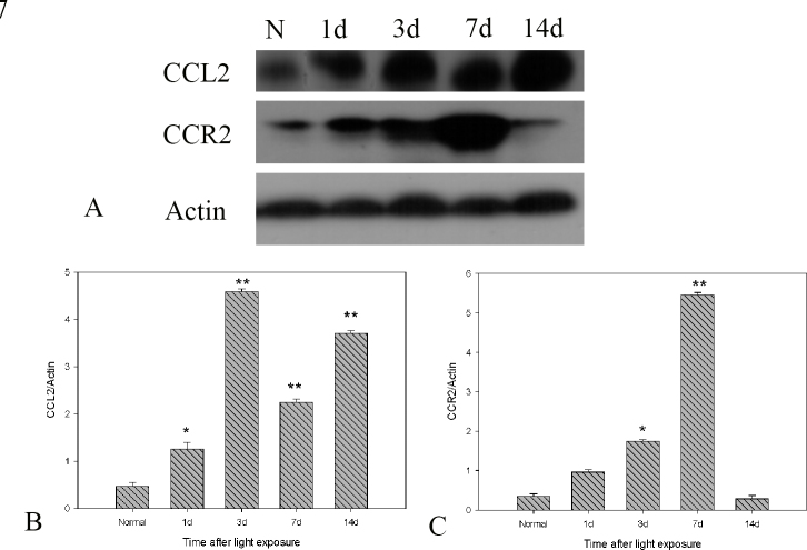
Real-time PCR analyses showed that CCL2 and CCR2 expression gradually increased after light exposure and peaked at 3 days, coinciding with the infiltration of OX42-positive cells and the expression of IL-1 beta and TNF-alpha in the outer retina. Double immunostaining of CCL2 with TUNEL revealed that CCL2 was expressed robustly in about 30% of the apoptotic photoreceptors at the early stage. As degeneration progressed, immunostaining of CCL2 with OX42 showed that activated and migrated microglia and monocytes expressed CCL2. At the late stage, Müller cells became the main source of CCL2, which was illustrated by CCL2 immunostaining with GFAP.
Light exposure led to apoptosis of photoreceptors, which expressed CCL2, accelerating an inflammation-mediated cascade by activating and attracting microglia and monocytes and promoting their secretion of CCL2 in the injured position.
探讨CCL2和CCR2系统在光诱导的光感受器细胞凋亡过程中对小胶质细胞和单核细胞激活及迁移的影响。
在光照后1天、3天、7天和14天,采用OX42和ED1免疫染色标记小胶质细胞和单核细胞的激活及迁移情况。应用CCL2与末端脱氧核苷酸转移酶介导的dUTP缺口末端标记法(TUNEL)、OX42或胶质纤维酸性蛋白(GFAP)进行双重免疫染色,以探究CCL2、凋亡光感受器、激活的小胶质细胞和单核细胞以及大胶质细胞(穆勒细胞和星形胶质细胞)之间的关系。采用实时聚合酶链反应(PCR)评估视网膜中CCL2和CCR2以及促炎因子白细胞介素(IL)-1β和肿瘤坏死因子(TNF)-α的mRNA水平。
实时PCR分析显示,光照后CCL2和CCR2表达逐渐增加,并在3天时达到峰值,这与OX42阳性细胞在外核层的浸润以及IL-1β和TNF-α在外核层的表达时间一致。CCL2与TUNEL的双重免疫染色显示,在早期约30%的凋亡光感受器中CCL2表达强烈。随着变性进展,CCL2与OX42的免疫染色显示,激活并迁移的小胶质细胞和单核细胞表达CCL2。在晚期,穆勒细胞成为CCL第二的主要来源,这通过CCL2与GFAP的免疫染色得以证实。
光照导致表达CCL2的光感受器细胞凋亡,通过激活并吸引小胶质细胞和单核细胞以及促进它们在损伤部位分泌CCL2,加速炎症介导的级联反应。