Hecht Silke, Anderson Kimberly M, Castel Aude, Griffin John F, Hespel Adrien-Maxence, Nelson Nathan, Sun Xiaocun
Department of Small Animal Clinical Sciences, University of Tennessee, Knoxville, TN, United States.
Department of Large Animal Clinical Sciences, College of Veterinary Medicine and Biomedical Sciences, Texas A&M University, College Station, TX, United States.
Front Vet Sci. 2021 Apr 22;8:603775. doi: 10.3389/fvets.2021.603775. eCollection 2021.
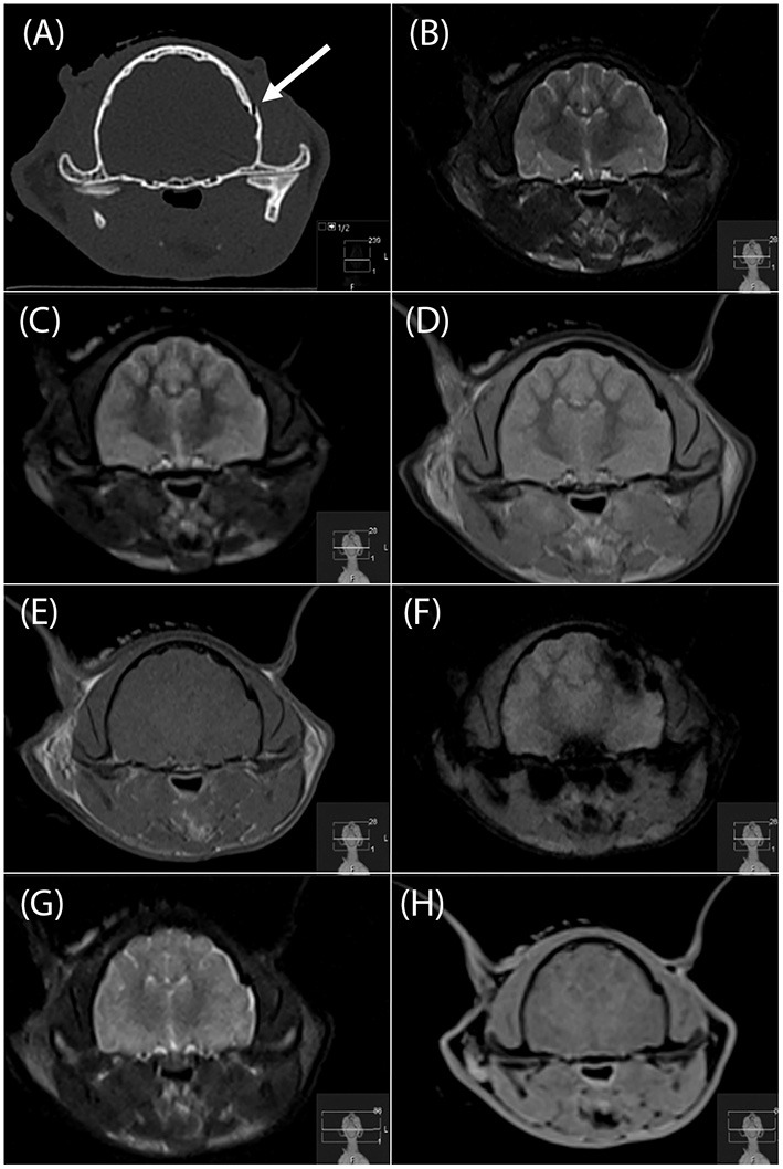
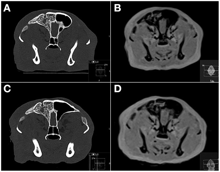
Computed tomography (CT) is the imaging modality of choice to evaluate patients with acute head trauma. However, magnetic resonance imaging (MRI) may be chosen in select cases. The objectives of this study were to evaluate the agreement of MRI with CT in the assessment for presence or absence of acute skull fractures in a canine and feline cadaver model, compare seven different MRI sequences (T1-W, T2-W, T2-FLAIR, PD-W, T2-W, "SPACE" and "VIBE"), and determine agreement of four different MRI readers with CT data. Pre- and post-trauma CT and MRI studies were performed on 10 canine and 10 feline cadaver heads. Agreement of MRI with CT as to presence or absence of a fracture was determined for 26 individual osseous structures and four anatomic regions (cranium, face, skull base, temporomandibular joint). Overall, there was 93.5% agreement in assessing a fracture as present or absent between MRI and CT, with a significant difference between the pre and post trauma studies (99.4 vs. 87.6%; < 0.0001; OR 0.042; 95% CI 0.034-0.052). There was no significant difference between dogs and cats. The agreement for the different MRI sequences with CT ranged from 92.6% (T2-W) to 94.4% (PD-W). There was higher agreement of MRI with CT in the evaluation for fractures of the face than other anatomic regions. Agreement with CT for individual MRI readers ranged from 92.6 to 94.7%. A PD-W sequence should be added to the MR protocol when evaluating the small animal head trauma patient.
计算机断层扫描(CT)是评估急性颅脑外伤患者的首选成像方式。然而,在某些特定情况下可选择磁共振成像(MRI)。本研究的目的是在犬猫尸体模型中评估MRI与CT在急性颅骨骨折有无评估方面的一致性,比较七种不同的MRI序列(T1加权、T2加权、T2液体衰减反转恢复序列、质子密度加权、T2加权、稳态采集空间谐波序列和容积内插屏气检查序列),并确定四位不同的MRI阅片者与CT数据的一致性。对10个犬类和10个猫类尸体头部进行创伤前后的CT和MRI研究。针对26个个体骨结构和四个解剖区域(颅骨、面部、颅底、颞下颌关节)确定MRI与CT在骨折有无方面的一致性。总体而言,MRI与CT在评估骨折有无方面的一致性为93.5%,创伤前后研究之间存在显著差异(99.4%对87.6%;P<0.0001;优势比0.042;95%置信区间0.034 - 0.052)。犬类和猫类之间无显著差异。不同MRI序列与CT的一致性范围为92.6%(T2加权)至94.4%(质子密度加权)。MRI与CT在面部骨折评估方面的一致性高于其他解剖区域。个体MRI阅片者与CT的一致性范围为92.6%至94.7%。在评估小动物头部外伤患者时,应在MR检查方案中添加质子密度加权序列。