Department of Botany, Functional Plant Biology, University of Innsbruck, 6020, Innsbruck, Austria.
Department of Plant and Environmental Sciences, Section for Plant Glycobiology, University of Copenhagen, 1871, Frederiksberg, Denmark.
Protoplasma. 2021 Nov;258(6):1261-1275. doi: 10.1007/s00709-021-01659-5. Epub 2021 May 11.
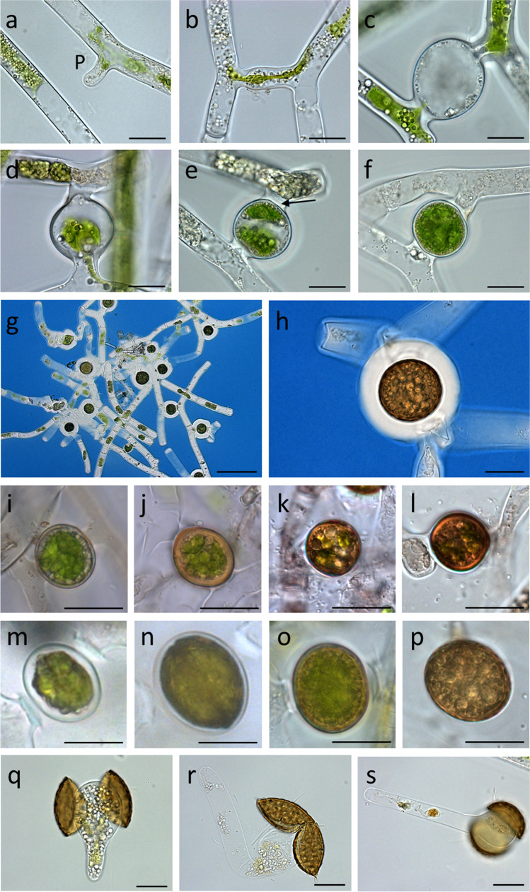
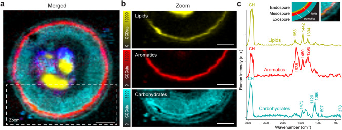
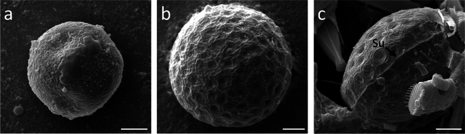
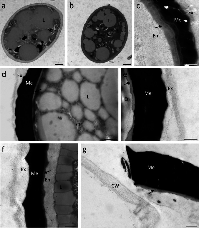
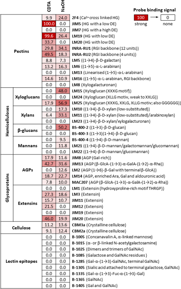
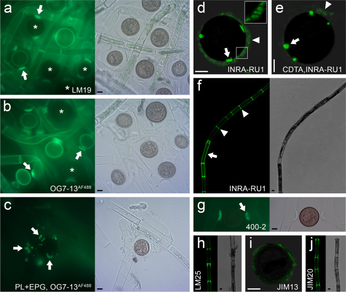
Mougeotia spp. collected from field samples were investigated for their conjugation morphology by light-, fluorescence-, scanning- and transmission electron microscopy. During a scalarifom conjugation, the extragametangial zygospores were initially surrounded by a thin cell wall that developed into a multi-layered zygospore wall. Maturing zygospores turned dark brown and were filled with storage compounds such as lipids and starch. While M. parvula had a smooth surface, M. disjuncta had a punctated surface structure and a prominent suture. The zygospore wall consisted of a polysaccharide rich endospore, followed by a thin layer with a lipid-like appaerance, a massive electron dense mesospore and a very thin exospore composed of polysaccharides. Glycan microarray analysis of zygospores of different developmental stages revealed the occurrence of pectins and hemicelluloses, mostly composed of homogalacturonan (HG), xyloglucans, xylans, arabino-galactan proteins and extensins. In situ localization by the probe OG7-13 labelled HG in young zygospore walls, vegetative filaments and most prominently in conjugation tubes and cross walls. Raman imaging showed the distribution of proteins, lipids, carbohydrates and aromatic components of the mature zygospore with a spatial resolution of ~ 250 nm. The carbohydrate nature of the endo- and exospore was confirmed and in-between an enrichment of lipids and aromatic components, probably algaenan or a sporopollenin-like material. Taken together, these results indicate that during zygospore formation, reorganizations of the cell walls occured, leading to a resistant and protective structure.
从野外样本中收集的 Mougeotia spp. 被用于通过光镜、荧光显微镜、扫描电子显微镜和透射电子显微镜研究其接合形态。在一个Scalarifom 接合过程中,外配子囊中的合子孢子最初被一层薄细胞壁包围,然后细胞壁逐渐发展成多层合子孢子壁。成熟的合子孢子变成深褐色,充满了脂质和淀粉等储存化合物。M. parvula 表面光滑,而 M. disjuncta 表面有斑点结构和明显的缝线。合子孢子壁由富含多糖的内孢子组成,其次是一层具有类脂质外观的薄层,一层密集的电子致密中孢子和一层由多糖组成的非常薄的外孢子。对不同发育阶段的合子孢子进行糖胺聚糖微阵列分析表明,果胶和半纤维素的存在,主要由同质半乳糖醛酸聚糖 (HG)、木葡聚糖、木聚糖、阿拉伯半乳聚糖蛋白和伸展蛋白组成。通过探针 OG7-13 进行的原位定位显示,HG 在年轻的合子孢子壁、营养丝和最明显的接合管和交叉壁中被标记。拉曼成像显示了成熟合子孢子的蛋白质、脂质、碳水化合物和芳香成分的分布,具有约 250nm 的空间分辨率。内孢子和外孢子的碳水化合物性质得到了确认,在脂质和芳香成分之间存在丰富的物质,可能是藻烯或类似于孢粉素的物质。总之,这些结果表明,在合子孢子形成过程中,细胞壁发生了重组,形成了一种具有抗性和保护结构的合子孢子。