Chen Chun, McDonald David, Blain Alasdair, Sachdeva Ashwin, Bone Laura, Smith Anna L M, Warren Charlotte, Pickett Sarah J, Hudson Gavin, Filby Andrew, Vincent Amy E, Turnbull Doug M, Reeve Amy K
Wellcome Centre for Mitochondrial Research, Translational and Clinical Research Institute, Newcastle University, Newcastle upon Tyne, UK.
Wellcome Centre for Mitochondrial Research, Biosciences Institute, Newcastle University, Newcastle upon Tyne, UK.
NPJ Parkinsons Dis. 2021 May 12;7(1):39. doi: 10.1038/s41531-021-00182-x.
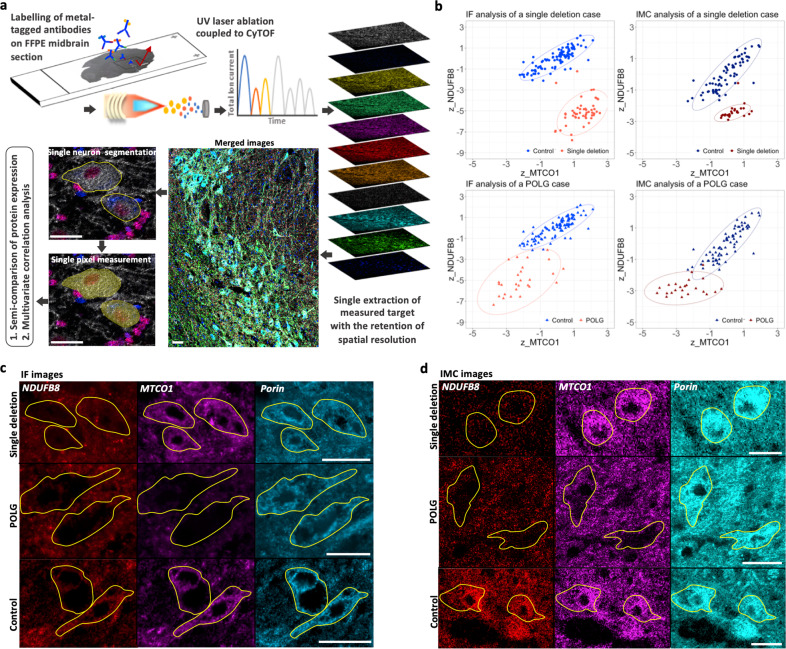
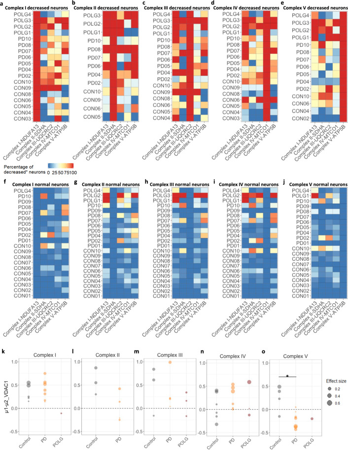
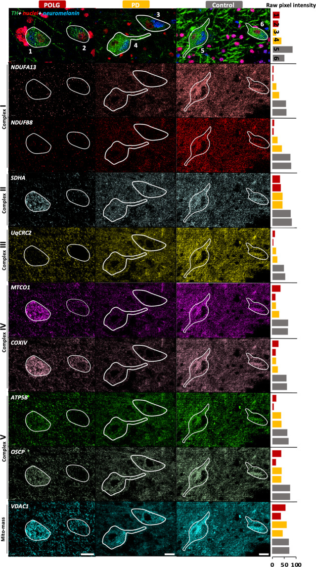
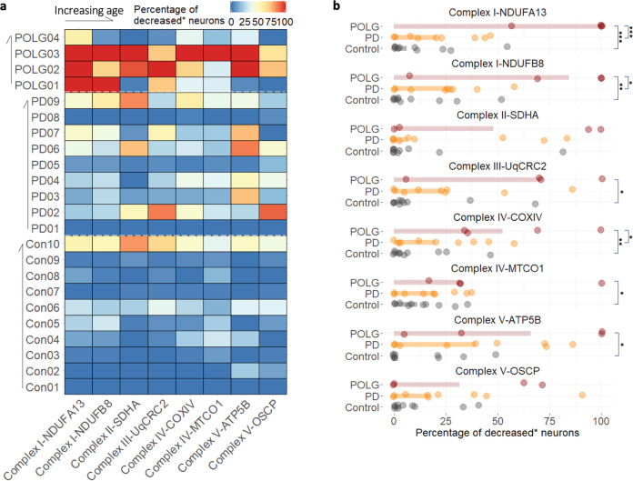
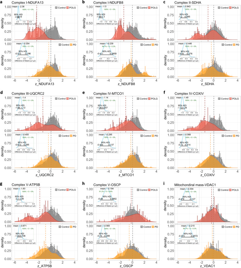
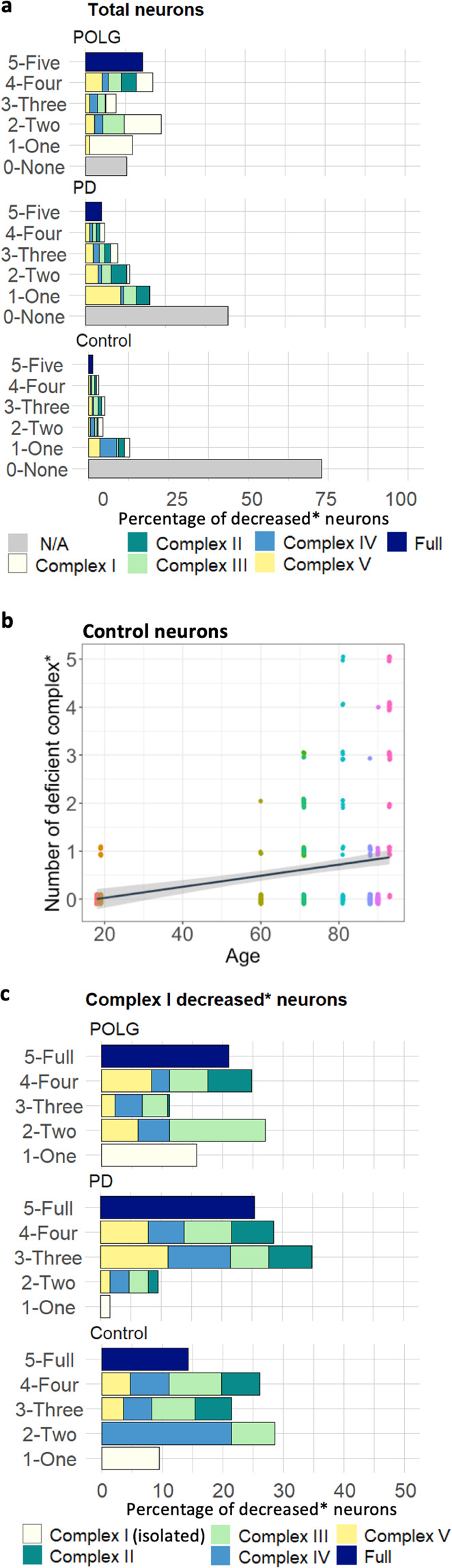
Here we report the application of a mass spectrometry-based technology, imaging mass cytometry, to perform in-depth proteomic profiling of mitochondrial complexes in single neurons, using metal-conjugated antibodies to label post-mortem human midbrain sections. Mitochondrial dysfunction, particularly deficiency in complex I has previously been associated with the degeneration of dopaminergic neurons in Parkinson's disease. To further our understanding of the nature of this dysfunction, and to identify Parkinson's disease specific changes, we validated a panel of antibodies targeting subunits of all five mitochondrial oxidative phosphorylation complexes in dopaminergic neurons from Parkinson's disease, mitochondrial disease, and control cases. Detailed analysis of the expression profile of these proteins, highlighted heterogeneity between individuals. There is a widespread decrease in expression of all complexes in Parkinson's neurons, although more severe in mitochondrial disease neurons, however, the combination of affected complexes varies between the two groups. We also provide evidence of a potential neuronal response to mitochondrial dysfunction through a compensatory increase in mitochondrial mass. This study highlights the use of imaging mass cytometry in the assessment and analysis of expression of oxidative phosphorylation proteins, revealing the complexity of deficiencies of these proteins within individual neurons which may contribute to and drive neurodegeneration in Parkinson's disease.
在此,我们报告了一种基于质谱的技术——成像质谱流式细胞术的应用,该技术使用金属偶联抗体标记死后人类中脑切片,对单个神经元中的线粒体复合物进行深入的蛋白质组分析。线粒体功能障碍,尤其是复合体I的缺陷,此前一直与帕金森病中多巴胺能神经元的退化有关。为了进一步了解这种功能障碍的本质,并识别帕金森病的特异性变化,我们验证了一组针对帕金森病、线粒体疾病和对照病例中多巴胺能神经元所有五个线粒体氧化磷酸化复合体亚基的抗体。对这些蛋白质表达谱的详细分析突出了个体之间的异质性。帕金森病神经元中所有复合体的表达普遍下降,尽管在线粒体疾病神经元中更严重,然而,两组中受影响复合体的组合有所不同。我们还提供了证据,表明通过线粒体质量的代偿性增加,神经元对线粒体功能障碍可能存在潜在反应。这项研究强调了成像质谱流式细胞术在评估和分析氧化磷酸化蛋白表达方面的应用,揭示了单个神经元中这些蛋白缺陷的复杂性,这可能导致并推动帕金森病中的神经退行性变。