Kummari Evangel, Guo-Ross Shirley X, Partington Heath S, Nutter Jennifer Makenzie, Eells Jeffrey B
Department of Biology, Texas A&M University, College Station, TX, United States.
Department of Basic Sciences, College of Veterinary Medicine, Mississippi State University, Starkville, MS, United States.
Front Neuroanat. 2021 Apr 30;15:563854. doi: 10.3389/fnana.2021.563854. eCollection 2021.
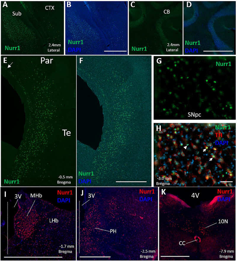
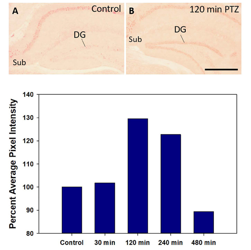
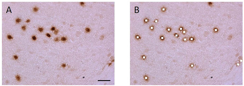
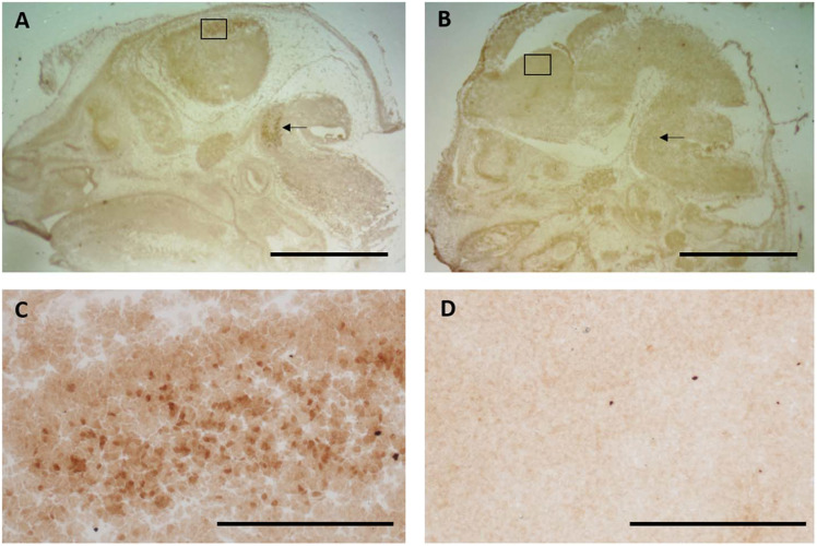
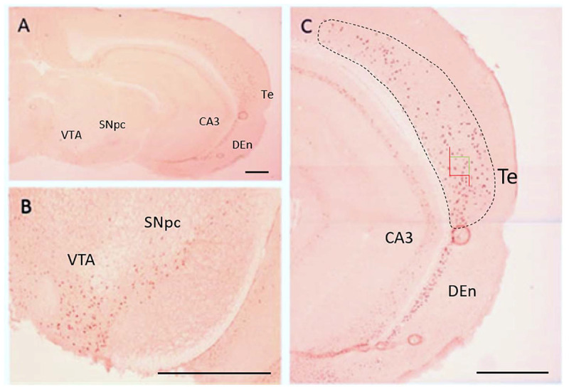
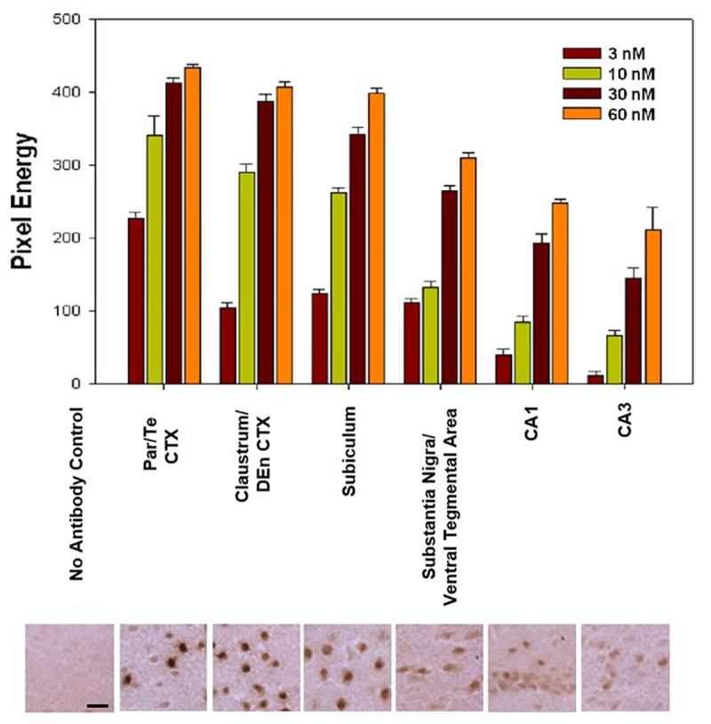
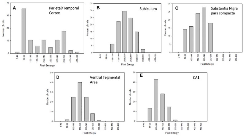
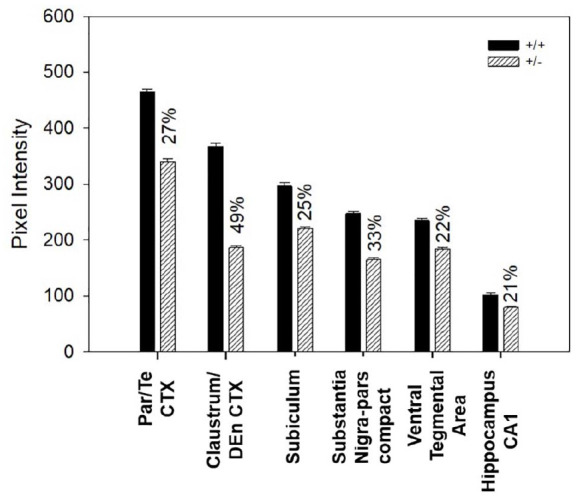
The transcription factor Nurr1 is a member of the steroid hormone nuclear receptor superfamily. Ablation of Nurr1 expression arrests mesencephalic dopamine neuron differentiation while attenuation of Nurr1 in the subiculum and hippocampus impairs learning and memory. Additionally, reduced Nurr1 expression has been reported in patients with Parkinson's disease and Alzheimer's disease. In order to better understand the overall function of Nurr1 in the brain, quantitative immunohistochemistry was used to measure cellular Nurr1 protein expression, across Nurr1 immunoreactive neuronal populations. Additionally, neuronal Nurr1 expression levels were compared between different brain regions in wild-type mice (+/+) and Nurr1 heterozygous mice (+/-). Regional Nurr1 protein was also investigated at various time points after a seizure induced by pentylenetetrazol (PTZ). Nurr1 protein is expressed in various regions throughout the brain, however, a wide range of Nurr1 expression levels were observed among various neuronal populations. Neurons in the parietal and temporal cortex (secondary somatosensory, insular, auditory, and temporal association cortex) had the highest relative Nurr1 expression (100%) followed closely by the claustrum/dorsal endopiriform cortex (85%) and then subiculum (76%). Lower Nurr1 protein levels were found in neurons in the substantia nigra pars compacta and ventral tegmental area (39%) followed by CA1 (25%) and CA3 (19%) of the hippocampus. Additionally, in the parietal and temporal cortex, two distinct populations of high and medium Nurr1 expressing neurons were observed. Comparisons between +/- and +/+ mice revealed Nurr1 protein was reduced in +/- mice by 27% in the parietal/temporal cortex, 49% in the claustrum/dorsal endopiriform cortex, 25% in the subiculum, 33% in substantia nigra pars compacta, 22% in ventral tegmental area, and 21% in CA1 region of the hippocampus. Based on these data, regional mechanisms appear to exist which can compensate for a loss of a Nurr1 allele. Following a single PTZ-induced seizure, Nurr1 protein in the dentate gyrus peaked around 2 h and returned to baseline by 8 h. Since altered Nurr1 expression has been implicated in neurologic disorders and Nurr1 agonists have showed protective effects, understanding regional protein expression of Nurr1, therefore, is necessary to understand how changes in Nurr1 expression can alter brain function.
转录因子Nurr1是类固醇激素核受体超家族的成员。Nurr1表达缺失会阻止中脑多巴胺神经元分化,而海马下托和海马中Nurr1表达减弱会损害学习和记忆。此外,据报道帕金森病和阿尔茨海默病患者的Nurr1表达降低。为了更好地了解Nurr1在大脑中的整体功能,采用定量免疫组织化学方法检测了Nurr1免疫反应性神经元群体中的细胞Nurr1蛋白表达。此外,还比较了野生型小鼠(+/+)和Nurr1杂合小鼠(+/-)不同脑区的神经元Nurr1表达水平。还研究了戊四氮(PTZ)诱导癫痫发作后不同时间点的脑区Nurr1蛋白。Nurr1蛋白在全脑的各个区域均有表达,然而,在不同的神经元群体中观察到Nurr1表达水平差异很大。顶叶和颞叶皮质(次级体感、岛叶、听觉和颞叶联合皮质)的神经元Nurr1相对表达最高(100%),紧随其后的是屏状核/背侧梨状内皮层(85%),然后是海马下托(76%)。黑质致密部和腹侧被盖区的神经元Nurr1蛋白水平较低(39%),其次是海马的CA1区(25%)和CA3区(19%)。此外,在顶叶和颞叶皮质中,观察到两个不同的高表达和中等表达Nurr1的神经元群体。+/ - 小鼠和+/ +小鼠的比较显示,+/ - 小鼠顶叶/颞叶皮质中的Nurr1蛋白减少27%,屏状核/背侧梨状内皮层减少49%,海马下托减少25%,黑质致密部减少33%,腹侧被盖区减少22%,海马CA1区减少21%。基于这些数据,似乎存在区域机制可以补偿Nurr1等位基因的缺失。单次PTZ诱导癫痫发作后,齿状回中的Nurr1蛋白在约2小时达到峰值,并在8小时恢复到基线水平。由于Nurr1表达改变与神经系统疾病有关,且Nurr1激动剂已显示出保护作用,但理解Nurr1的区域蛋白表达对于了解Nurr1表达变化如何改变脑功能是必要的。