Department of Neurology, Memory and Aging Center, Weill Institute for Neurosciences, University of California, San Francisco, 675 Nelson Rising Lane, Suite 190, San Francisco, CA, 94143, USA.
Departments of Neurology, Radiology & Biomedical Imaging, Memory and Aging Center, Weill Institute for Neurosciences, University of California, San Francisco, 675 Nelson Rising Lane, Suite 190, San Francisco, CA, 94143, USA.
Acta Neuropathol Commun. 2021 May 22;9(1):96. doi: 10.1186/s40478-021-01197-4.
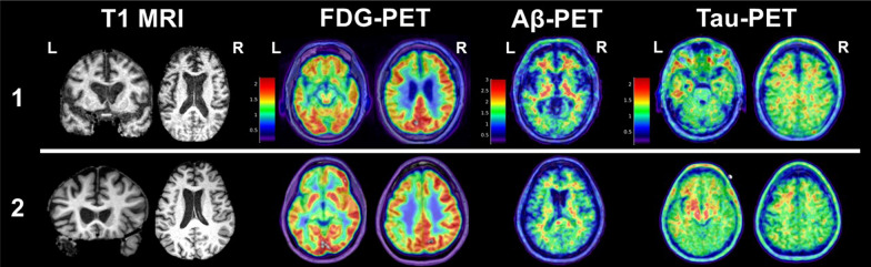
Varying severities and frequencies of head trauma may result in dynamic acute and chronic pathophysiologic responses in the brain. Heightened attention to long-term effects of head trauma, particularly repetitive head trauma, has sparked recent efforts to identify neuroimaging biomarkers of underlying disease processes. Imaging modalities like structural magnetic resonance imaging (MRI) and positron emission tomography (PET) are the most clinically applicable given their use in neurodegenerative disease diagnosis and differentiation. In recent years, researchers have targeted repetitive head trauma cohorts in hopes of identifying in vivo biomarkers for underlying biologic changes that might ultimately improve diagnosis of chronic traumatic encephalopathy (CTE) in living persons. These populations most often include collision sport athletes (e.g., American football, boxing) and military veterans with repetitive low-level blast exposure. We provide a clinically-oriented review of neuroimaging data from repetitive head trauma cohorts based on structural MRI, FDG-PET, Aβ-PET, and tau-PET. We supplement the review with two patient reports of neuropathology-confirmed, clinically impaired adults with prior repetitive head trauma who underwent structural MRI, FDG-PET, Aβ-PET, and tau-PET in addition to comprehensive clinical examinations before death.
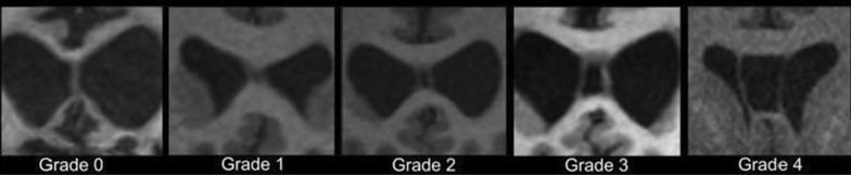
Group-level comparisons to controls without known head trauma have revealed inconsistent regional volume differences, with possible propensity for medial temporal, limbic, and subcortical (thalamus, corpus callosum) structures. Greater frequency and severity (i.e., length) of cavum septum pellucidum (CSP) is observed in repetitive head trauma cohorts compared to unexposed controls. It remains unclear whether CSP predicts a particular neurodegenerative process, but CSP presence should increase suspicion that clinical impairment is at least partly attributable to the individual's head trauma exposure (regardless of underlying disease). PET imaging similarly has not revealed a prototypical metabolic or molecular pattern associated with repetitive head trauma or predictive of CTE based on the most widely studied radiotracers. Given the range of clinical syndromes and neurodegenerative pathologies observed in a subset of adults with prior repetitive head trauma, structural MRI and PET imaging may still be useful for differential diagnosis (e.g., assessing suspected Alzheimer's disease).
头部创伤的严重程度和频率不同,可能导致大脑出现动态的急性和慢性病理生理反应。人们越来越关注头部创伤的长期影响,特别是重复性头部创伤,这激发了最近的努力,以确定潜在疾病过程的神经影像学生物标志物。鉴于其在神经退行性疾病诊断和鉴别中的应用,像结构磁共振成像(MRI)和正电子发射断层扫描(PET)这样的成像方式是最具临床适用性的。近年来,研究人员针对重复性头部创伤队列进行了研究,希望能够确定潜在生物学变化的体内生物标志物,这可能最终有助于在活体中诊断慢性创伤性脑病(CTE)。这些人群通常包括碰撞运动运动员(如美式足球、拳击)和反复受到低水平爆炸冲击的退伍军人。我们提供了基于结构 MRI、FDG-PET、Aβ-PET 和 tau-PET 的重复性头部创伤队列的神经影像学数据的临床导向综述。我们补充了两个患者报告,这些患者生前有过重复性头部创伤,经神经病理学证实存在认知障碍,除了全面的临床检查外,他们在死亡前还接受了结构 MRI、FDG-PET、Aβ-PET 和 tau-PET。
与无已知头部创伤的对照组进行的组间比较显示,区域体积存在不一致的差异,可能倾向于内侧颞叶、边缘和皮质下(丘脑、胼胝体)结构。与未暴露于创伤的对照组相比,重复性头部创伤队列中观察到更频繁和更严重(即更长)的透明隔腔(CSP)。CSP 是否预示着特定的神经退行性过程尚不清楚,但 CSP 的存在应该增加对认知障碍至少部分归因于个体头部创伤暴露的怀疑(无论潜在疾病如何)。PET 成像同样没有显示出与重复性头部创伤相关的典型代谢或分子模式,也没有根据最广泛研究的示踪剂预测 CTE。鉴于一组先前有过重复性头部创伤的成年人中观察到的一系列临床综合征和神经退行性病理,结构 MRI 和 PET 成像可能仍然有助于鉴别诊断(例如,评估疑似阿尔茨海默病)。