Department of Neurosurgery, Iwate Medical University School of Medicine, Yahaba, Japan.
Department of Neurology and Gerontology, Iwate Medical University School of Medicine, Yahaba, Japan.
Cerebrovasc Dis Extra. 2021;11(2):61-68. doi: 10.1159/000516426. Epub 2021 May 25.
During exposure of the carotid arteries, embolism from the surgical site is recognized as a primary cause of neurological deficits or new cerebral ischemic lesions following carotid endarterectomy (CEA), and associations have been reported between histological neovascularization in the carotid plaque and both plaque vulnerability and the development of artery-to-artery embolism. Superb microvascular imaging (SMI) enables accurate visualization of neovessels in the carotid plaque without the use of intravenous contrast. This study aimed to determine whether preoperative SMI ultrasound for cervical carotid artery stenosis predicts the development of microembolic signals (MES) on transcranial Doppler (TCD) during exposure of the carotid arteries in CEA.
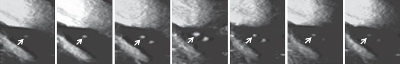
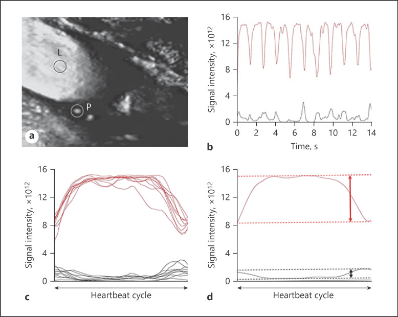
Preoperative cervical carotid artery SMI ultrasound followed by CEA under TCD monitoring of MES in the ipsilateral middle cerebral artery was conducted in 70 patients previously diagnosed with internal carotid artery stenosis (defined as ≥70%). First, observers visually identified intraplaque microvascular flow (IMVF) signals as moving enhancements located near the surface of the carotid plaque within the plaque on SMI ultrasonograms. Next, regions of interest (ROI) were manually placed at the identified IMVF signals (or at arbitrary places within the plaque when no IMVF signals were identified within the carotid plaque) and the carotid lumen, and time-intensity curves of the IMVF signal and lumen ROI were generated. Ten heartbeat cycles of both time-intensity curves were segmented into each heartbeat cycle based on gated electrocardiogram findings and averaged with respect to the IMVF signal and lumen ROI. The difference between the maximum and minimum intensities (ID) was calculated based on the averaged IMVF signal (IDIMVF) and lumen (IDl) curves. Finally, the ratio of IDIMVF to IDl was calculated.
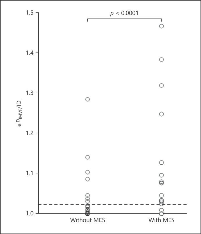
MES during exposure of the carotid arteries were detected in 17 patients (24%). The incidence of identification of IMVF signals was significantly greater in patients with MES (94%) than in those without (57%; p = 0.0067). The IDIMVF/IDl ratio was significantly greater in patients with MES (0.108 ± 0.120) than in those without (0.017 ± 0.042; p < 0.0001). The specificity and positive predictive value for the IDIMVF/IDl ratio for prediction of the development of MES were significantly higher than those for the identification of IMVF signals. Logistic regression analysis revealed that only the IDIMVF/IDl ratio was significantly associated with the development of MES (95% CI 101.1-3,628.9; p = 0.0048).
Preoperative cervical carotid artery SMI ultrasound predicts the development of MES on TCD during exposure of the carotid arteries in CEA.
在颈动脉暴露过程中,手术部位的栓塞被认为是颈动脉内膜切除术(CEA)后发生神经功能缺损或新的脑缺血性病变的主要原因,颈动脉斑块内的组织新生血管化与斑块易损性和动脉到动脉栓塞的发展之间存在关联。卓越的微血管成像(SMI)可在不使用静脉内对比的情况下准确显示颈动脉斑块内的新生血管。本研究旨在确定术前颈内颈动脉 SMI 超声是否可预测 CEA 中颈动脉暴露时经颅多普勒(TCD)上微栓子信号(MES)的发展。
70 例先前诊断为颈内动脉狭窄(定义为≥70%)的患者进行了术前颈内颈动脉 SMI 超声检查,然后在 TCD 监测下进行 MES 的 CEA。首先,观察者在 SMI 超声图像上通过视觉识别斑块内的微血管血流(IMVF)信号,这些信号为位于颈动脉斑块表面附近的移动增强信号。然后,手动将感兴趣区(ROI)置于识别的 IMVF 信号处(或在颈动脉斑块内未识别到 IMVF 信号的情况下置于斑块内的任意位置),并生成 IMVF 信号和管腔 ROI 的时间强度曲线。根据门控心电图检查结果,将 10 个心跳周期的两个时间强度曲线分段为每个心跳周期,并分别对 IMVF 信号和管腔 ROI 进行平均处理。根据平均 IMVF 信号(IDIMVF)和管腔(IDl)曲线计算最大和最小强度之间的差异(ID)。最后,计算 IDIMVF 与 IDl 的比值。
在 17 名患者(24%)中检测到颈动脉暴露时的 MES。有 MES 的患者识别出 IMVF 信号的发生率明显高于无 MES 的患者(94% vs. 57%;p=0.0067)。有 MES 的患者的 IDIMVF/IDl 比值明显大于无 MES 的患者(0.108±0.120 vs. 0.017±0.042;p<0.0001)。对于预测 MES 的发展,IDIMVF/IDl 比值的特异性和阳性预测值明显高于识别 IMVF 信号的特异性和阳性预测值。Logistic 回归分析显示,只有 IDIMVF/IDl 比值与 MES 的发展显著相关(95%CI 101.1-3628.9;p=0.0048)。
术前颈内颈动脉 SMI 超声可预测 CEA 中颈动脉暴露时 TCD 上 MES 的发生。