• 文献检索
  • 文档翻译
  • 深度研究
  • 学术资讯
  • Suppr Zotero 插件Zotero 插件
  • 邀请有礼
  • 套餐&价格
  • 历史记录
应用&插件
Suppr Zotero 插件Zotero 插件浏览器插件Mac 客户端Windows 客户端微信小程序
定价
高级版会员购买积分包购买API积分包
服务
文献检索文档翻译深度研究API 文档MCP 服务
关于我们
关于 Suppr公司介绍联系我们用户协议隐私条款
关注我们

Suppr 超能文献

核心技术专利:CN118964589B侵权必究
粤ICP备2023148730 号-1Suppr @ 2026

文献检索

告别复杂PubMed语法,用中文像聊天一样搜索,搜遍4000万医学文献。AI智能推荐,让科研检索更轻松。

立即免费搜索

文件翻译

保留排版,准确专业,支持PDF/Word/PPT等文件格式,支持 12+语言互译。

免费翻译文档

深度研究

AI帮你快速写综述,25分钟生成高质量综述,智能提取关键信息,辅助科研写作。

立即免费体验

丹参酮 I 通过 JNK/ERK 介导的凋亡通路抑制人慢性髓系白血病细胞的生长。

Tanshinone I inhibited growth of human chronic myeloid leukemia cells via JNK/ERK mediated apoptotic pathways.

机构信息

The First Affiliated Hospital of Zhejiang Chinese Medical University, Hangzhou, China.

College of Pharmaceutical Science, Zhejiang Chinese Medical University, Hangzhou, China.

出版信息

Braz J Med Biol Res. 2021 May 24;54(8):e10685. doi: 10.1590/1414-431X2020e10685. eCollection 2021.

DOI:10.1590/1414-431X2020e10685
PMID:34037092
原文链接:https://pmc.ncbi.nlm.nih.gov/articles/PMC8148979/
Abstract

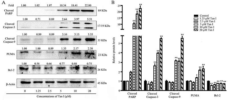
Tanshinone I (Tan I) is one of the main bioactive ingredients derived from Salvia miltiorrhiza Bunge, which has exhibited antitumor activities toward various human cancer cells. However, its effects and underlying mechanisms on human chronic myeloid leukemia (CML) cells still require further investigation. This study determined the effects and mechanisms of anti-proliferative and apoptosis induction activity induced by Tan I against K562 cells. The cytotoxic effect of Tan I at varying concentrations on K562 cells was evaluated via MTT assay. Cell apoptosis was further investigated through DAPI staining and flow cytometry analysis. The expression levels of apoptosis-related proteins and activities of JNK/ATF2 and ERK signaling pathways were analyzed by western blot. Quantitative PCR was performed to further determine mRNA expression levels of JNK1/2 and ERK1/2 after Tan I treatment. The results indicated that Tan I significantly inhibited K562 cell growth and induced apoptosis in a concentration- and time-dependent manner. It induced significant cellular morphological changes and increased apoptosis rates in CML cells. Tan I promoted the cleavages of caspase-related proteins, as well as increased the expression levels of PUMA. Furthermore, Tan I significantly activated JNK and inhibited ATF-2 and ERK signaling pathways. The mRNA expression levels of JNK1/2 and ERK1/2 were up-regulated by Tan I, further confirming its regulatory effects on JNK/ERK signaling pathways. Overall, our results indicated that Tan I suppressed cell viability via JNK- and ERK-mediated apoptotic pathways in K562 cells, suggesting that it might be a promising candidate as a novel anti-leukemia drug.

摘要

丹参酮 I(Tan I)是从丹参中提取的主要生物活性成分之一,对多种人类癌细胞表现出抗肿瘤活性。然而,其对人慢性髓系白血病(CML)细胞的作用及其机制仍需进一步研究。本研究旨在确定 Tan I 对 K562 细胞的抗增殖和诱导凋亡活性的作用及其机制。通过 MTT 法评估 Tan I 在不同浓度下对 K562 细胞的细胞毒性作用。通过 DAPI 染色和流式细胞术分析进一步研究细胞凋亡。通过 Western blot 分析凋亡相关蛋白的表达水平以及 JNK/ATF2 和 ERK 信号通路的活性。通过定量 PCR 进一步确定 Tan I 处理后 JNK1/2 和 ERK1/2 的 mRNA 表达水平。结果表明,Tan I 以浓度和时间依赖性方式显著抑制 K562 细胞生长并诱导细胞凋亡。它诱导 CML 细胞发生明显的细胞形态变化并增加细胞凋亡率。Tan I 促进了与 caspase 相关的蛋白的裂解,同时增加了 PUMA 的表达水平。此外,Tan I 显著激活了 JNK 并抑制了 ATF-2 和 ERK 信号通路。Tan I 上调了 JNK1/2 和 ERK1/2 的 mRNA 表达水平,进一步证实了其对 JNK/ERK 信号通路的调节作用。总之,我们的研究结果表明,Tan I 通过 JNK 和 ERK 介导的凋亡途径抑制 K562 细胞活力,表明其可能是一种有前途的新型抗白血病药物候选物。

https://cdn.ncbi.nlm.nih.gov/pmc/blobs/d50d/8148979/6966963a504b/1414-431X-bjmbr-54-8-e10685-gf006.jpg
https://cdn.ncbi.nlm.nih.gov/pmc/blobs/d50d/8148979/1e0a2e2b1939/1414-431X-bjmbr-54-8-e10685-gf001.jpg
https://cdn.ncbi.nlm.nih.gov/pmc/blobs/d50d/8148979/750cc7486bff/1414-431X-bjmbr-54-8-e10685-gf002.jpg
https://cdn.ncbi.nlm.nih.gov/pmc/blobs/d50d/8148979/de47f7db497f/1414-431X-bjmbr-54-8-e10685-gf003.jpg
https://cdn.ncbi.nlm.nih.gov/pmc/blobs/d50d/8148979/83a1f87d41f5/1414-431X-bjmbr-54-8-e10685-gf004.jpg
https://cdn.ncbi.nlm.nih.gov/pmc/blobs/d50d/8148979/32eb664303c0/1414-431X-bjmbr-54-8-e10685-gf005.jpg
https://cdn.ncbi.nlm.nih.gov/pmc/blobs/d50d/8148979/6966963a504b/1414-431X-bjmbr-54-8-e10685-gf006.jpg

相似文献

1
Tanshinone I inhibited growth of human chronic myeloid leukemia cells via JNK/ERK mediated apoptotic pathways.丹参酮 I 通过 JNK/ERK 介导的凋亡通路抑制人慢性髓系白血病细胞的生长。
Braz J Med Biol Res. 2021 May 24;54(8):e10685. doi: 10.1590/1414-431X2020e10685. eCollection 2021.
2
Down-regulation of telomerase activity and activation of caspase-3 are responsible for Tanshinone I-induced apoptosis in monocyte leukemia cells in vitro.端粒酶活性下调和半胱天冬酶-3激活是丹参酮I体外诱导单核细胞白血病细胞凋亡的原因。
Int J Mol Sci. 2010 May 26;11(6):2267-80. doi: 10.3390/ijms11062267.
3
Tanshinone I attenuates the malignant biological properties of ovarian cancer by inducing apoptosis and autophagy via the inactivation of PI3K/AKT/mTOR pathway.丹参酮 I 通过抑制 PI3K/AKT/mTOR 通路诱导细胞凋亡和自噬来抑制卵巢癌细胞的恶性生物学特性。
Cell Prolif. 2020 Feb;53(2):e12739. doi: 10.1111/cpr.12739. Epub 2019 Dec 9.
4
Analysis of tanshinone IIA induced cellular apoptosis in leukemia cells by genome-wide expression profiling.通过全基因组表达谱分析丹参酮 IIA 诱导白血病细胞凋亡。
BMC Complement Altern Med. 2012 Jan 16;12:5. doi: 10.1186/1472-6882-12-5.
5
Saponins from Rubus parvifolius L. induce apoptosis in human chronic myeloid leukemia cells through AMPK activation and STAT3 inhibition.悬钩子叶中的皂苷通过激活AMPK和抑制STAT3诱导人慢性粒细胞白血病细胞凋亡。
Asian Pac J Cancer Prev. 2014;15(13):5455-61. doi: 10.7314/apjcp.2014.15.13.5455.
6
BCR/ABL mRNA targeting small interfering RNA effects on proliferation and apoptosis in chronic myeloid leukemia.靶向BCR/ABL mRNA的小干扰RNA对慢性髓性白血病细胞增殖和凋亡的影响
Asian Pac J Cancer Prev. 2014;15(12):4773-80. doi: 10.7314/apjcp.2014.15.12.4773.
7
The Tanshinones (Tan) Extract From Bunge Induces ROS-Dependent Apoptosis in Pancreatic Cancer via AKT Hyperactivation-Mediated FOXO3/SOD2 Signaling.丹参酮(Tan)提取物通过 AKT 过度激活介导的 FOXO3/SOD2 信号诱导胰腺癌中的 ROS 依赖性细胞凋亡。
Integr Cancer Ther. 2024 Jan-Dec;23:15347354241258961. doi: 10.1177/15347354241258961.
8
Inactivation of PI3k/Akt signaling pathway and activation of caspase-3 are involved in tanshinone I-induced apoptosis in myeloid leukemia cells in vitro.丹参酮 I 诱导体外髓系白血病细胞凋亡涉及 PI3k/Akt 信号通路的失活和 caspase-3 的激活。
Ann Hematol. 2010 Nov;89(11):1089-97. doi: 10.1007/s00277-010-0996-z. Epub 2010 May 30.
9
Tanshinone IIA inhibits gastric carcinoma AGS cells through increasing p-p38, p-JNK and p53 but reducing p-ERK, CDC2 and cyclin B1 expression.丹参酮 IIA 通过增加 p-p38、p-JNK 和 p53,同时减少 p-ERK、CDC2 和细胞周期蛋白 B1 的表达来抑制胃癌 AGS 细胞。
Anticancer Res. 2014 Dec;34(12):7097-110.
10
Tanshinone IIA activates calcium-dependent apoptosis signaling pathway in human hepatoma cells.丹参酮 IIA 通过钙依赖性凋亡信号通路诱导人肝癌细胞凋亡。
J Nat Med. 2012 Jan;66(1):192-201. doi: 10.1007/s11418-011-0576-0. Epub 2011 Aug 19.

引用本文的文献

1
Tanshinone IIA and hepatocellular carcinoma: A potential therapeutic drug.丹参酮IIA与肝细胞癌:一种潜在的治疗药物。
Front Oncol. 2023 Jan 31;13:1071415. doi: 10.3389/fonc.2023.1071415. eCollection 2023.
2
in cancer: Potential role in regulating MicroRNAs and epigenetic enzymes.在癌症中:在调节微小RNA和表观遗传酶方面的潜在作用。
Front Pharmacol. 2022 Sep 12;13:1008222. doi: 10.3389/fphar.2022.1008222. eCollection 2022.

本文引用的文献

1
EM-2 inhibited autophagy and promoted G/M phase arrest and apoptosis by activating the JNK pathway in hepatocellular carcinoma cells.EM-2 通过激活 JNK 通路抑制自噬,促进肝癌细胞 G/M 期阻滞和凋亡。
Acta Pharmacol Sin. 2021 Jul;42(7):1139-1149. doi: 10.1038/s41401-020-00564-6. Epub 2020 Dec 14.
2
Saikosaponin D Inhibits Proliferation and Promotes Apoptosis Through Activation of MKK4-JNK Signaling Pathway in Pancreatic Cancer Cells.柴胡皂苷D通过激活MKK4-JNK信号通路抑制胰腺癌细胞增殖并促进其凋亡。
Onco Targets Ther. 2020 Sep 24;13:9465-9479. doi: 10.2147/OTT.S263322. eCollection 2020.
3
SULF2 promotes tumorigenesis and inhibits apoptosis of cervical cancer cells through the ERK/AKT signaling pathway.
SULF2 通过 ERK/AKT 信号通路促进宫颈癌的发生发展并抑制细胞凋亡。
Braz J Med Biol Res. 2020 Feb 7;53(2):e8901. doi: 10.1590/1414-431X20198901. eCollection 2020.
4
6'-O-galloylpaeoniflorin regulates proliferation and metastasis of non-small cell lung cancer through AMPK/miR-299-5p/ATF2 axis.6'-O-没食子酰基芍药苷通过 AMPK/miR-299-5p/ATF2 轴调控非小细胞肺癌的增殖和转移。
Respir Res. 2020 Feb 3;21(1):39. doi: 10.1186/s12931-020-1277-6.
5
Tanshinone I attenuates the malignant biological properties of ovarian cancer by inducing apoptosis and autophagy via the inactivation of PI3K/AKT/mTOR pathway.丹参酮 I 通过抑制 PI3K/AKT/mTOR 通路诱导细胞凋亡和自噬来抑制卵巢癌细胞的恶性生物学特性。
Cell Prolif. 2020 Feb;53(2):e12739. doi: 10.1111/cpr.12739. Epub 2019 Dec 9.
6
Tanshinone I inhibits the growth and metastasis of osteosarcoma via suppressing JAK/STAT3 signalling pathway.丹参酮 I 通过抑制 JAK/STAT3 信号通路抑制骨肉瘤的生长和转移。
J Cell Mol Med. 2019 Sep;23(9):6454-6465. doi: 10.1111/jcmm.14539. Epub 2019 Jul 10.
7
Molecular mechanism of miR-204 regulates proliferation, apoptosis and autophagy of cervical cancer cells by targeting ATF2.miR-204 通过靶向 ATF2 调控宫颈癌细胞增殖、凋亡及自噬的分子机制。
Artif Cells Nanomed Biotechnol. 2019 Dec;47(1):2529-2535. doi: 10.1080/21691401.2019.1628038.
8
Daucosterol induces autophagic-dependent apoptosis in prostate cancer via JNK activation.大根香叶醇通过激活 JNK 诱导前列腺癌细胞发生自噬依赖性凋亡。
Biosci Trends. 2019 May 12;13(2):160-167. doi: 10.5582/bst.2018.01293. Epub 2019 Apr 2.
9
Nuclear ERK: Mechanism of Translocation, Substrates, and Role in Cancer.核 ERK:易位机制、底物及在癌症中的作用。
Int J Mol Sci. 2019 Mar 8;20(5):1194. doi: 10.3390/ijms20051194.
10
Andrographolide and its potent derivative exhibit anticancer effects against imatinib-resistant chronic myeloid leukemia cells by downregulating the Bcr-Abl oncoprotein.穿心莲内酯及其有效衍生物通过下调 Bcr-Abl 癌蛋白对伊马替尼耐药的慢性髓系白血病细胞表现出抗癌作用。
Biochem Pharmacol. 2019 May;163:308-320. doi: 10.1016/j.bcp.2019.02.028. Epub 2019 Feb 26.