Kanazawa University Hospital, Respiratory Medicine, Ishikawa, Japan.
Kanazawa University, College of Medical, Pharmaceutical & Health Sciences, Ishikawa, Japan.
Int J Chron Obstruct Pulmon Dis. 2021 May 18;16:1393-1399. doi: 10.2147/COPD.S309960. eCollection 2021.
The aim of this study was to identify the relationships between parameters obtained from dynamic-ventilatory digital radiography (DR) and ventilatory disorders.
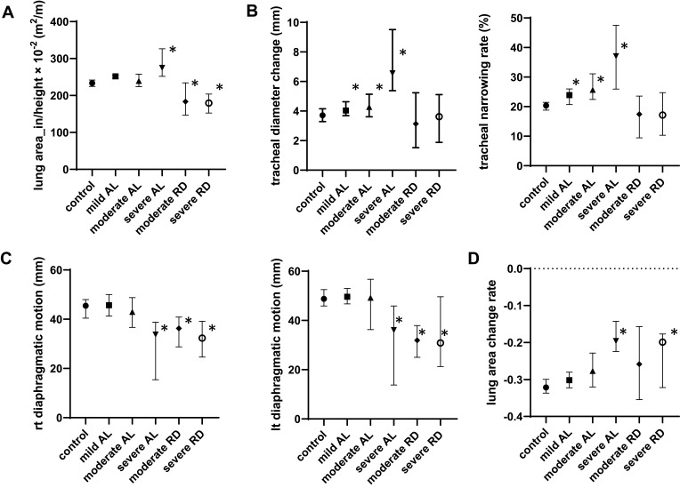
This study comprised 273 participants with respiratory diseases who underwent spirometry and functional residual capacity measurements (104 with normal findings on spirometry as controls, 139 with an obstructive lung disorder, 30 with a restrictive lung disorder) were assessed by dynamic-ventilatory DR. Sequential chest radiography images of the patient's slow and maximum breathing were captured at 15 frames per second by a dynamic flat-panel imaging system. The system measured the following parameters: lung area at maximum inspiration divided by height (lung area_in/height), changes in tracheal diameter due to respiratory motions, rate of tracheal narrowing, diaphragmatic motion, and rate of change in lung area due to respiratory motion. Relationships between these parameters and ventilatory disorders were analyzed.
Lung area_in/height in patients with restrictive disorders showed significant decreases. Tracheal diameter change and tracheal narrowing rate in patients with obstructive disorders were significantly increased compared to both the control participants and patients with restrictive disorders. Patients with obstructive disorders and patients with restrictive disorders showed decreased diaphragmatic motion and lung area change rate. With the restrictive disorders as references, the area under the curve (AUC), sensitivity and specificity of lung area_in/height were 0.88, 0.77, and 0.88, respectively. With the obstructive disorders as references, the AUC, sensitivity and specificity of tracheal narrowing rate were 0.67, 0.53 and 0.81, respectively.
Dynamic-ventilatory DR shows potential as a method for the detection and evaluation of ventilatory disorders in patients with respiratory diseases.
本研究旨在确定从动态通气数字 X 射线摄影(DR)获得的参数与通气障碍之间的关系。
本研究纳入了 273 名患有呼吸系统疾病的患者,他们接受了肺量计和功能残气量测量(104 名肺量计检查结果正常的患者作为对照组,139 名患有阻塞性肺疾病的患者,30 名患有限制性肺疾病的患者)。通过动态通气 DR 进行评估。动态平板成像系统以每秒 15 帧的速度连续拍摄患者缓慢和最大呼吸时的胸部 X 射线图像。该系统测量以下参数:最大吸气时的肺面积除以身高(肺面积 _in/height),由于呼吸运动导致的气管直径变化,气管变窄率,膈肌运动以及由于呼吸运动导致的肺面积变化率。分析了这些参数与通气障碍之间的关系。
限制性疾病患者的肺面积 _in/height 显著降低。与对照组和限制性疾病患者相比,阻塞性疾病患者的气管直径变化和气管狭窄率明显增加。阻塞性疾病患者和限制性疾病患者的膈肌运动和肺面积变化率均降低。以限制性疾病为参考,肺面积 _in/height 的曲线下面积(AUC)、灵敏度和特异性分别为 0.88、0.77 和 0.88。以阻塞性疾病为参考,气管狭窄率的 AUC、灵敏度和特异性分别为 0.67、0.53 和 0.81。
动态通气 DR 有望成为检测和评估呼吸系统疾病患者通气障碍的一种方法。