• 文献检索
  • 文档翻译
  • 深度研究
  • 学术资讯
  • Suppr Zotero 插件Zotero 插件
  • 邀请有礼
  • 套餐&价格
  • 历史记录
应用&插件
Suppr Zotero 插件Zotero 插件浏览器插件Mac 客户端Windows 客户端微信小程序
定价
高级版会员购买积分包购买API积分包
服务
文献检索文档翻译深度研究API 文档MCP 服务
关于我们
关于 Suppr公司介绍联系我们用户协议隐私条款
关注我们

Suppr 超能文献

核心技术专利:CN118964589B侵权必究
粤ICP备2023148730 号-1Suppr @ 2026

文献检索

告别复杂PubMed语法,用中文像聊天一样搜索,搜遍4000万医学文献。AI智能推荐,让科研检索更轻松。

立即免费搜索

文件翻译

保留排版,准确专业,支持PDF/Word/PPT等文件格式,支持 12+语言互译。

免费翻译文档

深度研究

AI帮你快速写综述,25分钟生成高质量综述,智能提取关键信息,辅助科研写作。

立即免费体验

肘部透视:一项尸体研究,确定用于显示重要解剖标志的新标准投照方法。

Fluoroscopy of the Elbow: A Cadaveric Study Defining New Standard Projections to Visualize Important Anatomical Landmarks.

作者信息

Müller Sebastian A, Adolfsson Lars, Baum Cornelia, Müller-Gerbl Magdalena, Müller Andreas M, Rikli Daniel

机构信息

Department of Orthopedic Surgery (S.A.M., C.B., A.M.M., and D.R.) and Institute of Anatomy (M.M.-G.), University of Basel, Basel, Switzerland.

Department of Orthopedic Surgery, Linköping University, Linköping, Sweden.

出版信息

JB JS Open Access. 2021 May 5;6(2). doi: 10.2106/JBJS.OA.20.00160. eCollection 2021 Apr-Jun.

DOI:10.2106/JBJS.OA.20.00160
PMID:34056508
原文链接:https://pmc.ncbi.nlm.nih.gov/articles/PMC8154458/
Abstract

BACKGROUND

Despite new 3-dimensional imaging modalities, 2-dimensional fluoroscopy remains the standard intraoperative imaging modality. The elbow has complex anatomy, and defined standard fluoroscopic projections are lacking. Therefore, the aim of this study was to define standard projections of the elbow for intraoperative fluoroscopy.

METHODS

This study consisted of 2 parts. In part I, dissected cadaveric elbows were examined under fluoroscopy, and their radiographic anatomical features were assessed, with focus on projections showing defined anatomical landmarks. In part II, projections from part I were verified on entire cadavers to simulate intraoperative imaging. Standard projections for anteroposterior (AP) and lateral views as well as oblique and axial views were recorded.

RESULTS

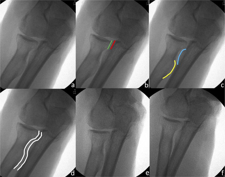
Eight standardized projections could be defined and included 3 AP, 1 lateral, 2 oblique, and 2 axial views. By applying these specific projections, we could visualize the epicondyles, the trochlea with its medial and lateral borders, the capitellum, the olecranon, the greater sigmoid notch, the coronoid process including its anteromedial facet, the proximal radioulnar joint with the radial tuberosity, and the anterior and posterior joint lines of the distal part of the humerus. These standard projections were reliably obtained using a specific sequence.

CONCLUSIONS

Knowledge about radiographic anatomy and standard projections is essential for visualizing important landmarks. With the presented standard projections of the elbow, important anatomical landmarks can be clearly examined. Thus, fluoroscopic visualization of anatomical fracture reduction and correct implant placement should be facilitated.

CLINICAL RELEVANCE

This basic science cadaveric study defines fluoroscopic standard projections of the elbow essential for visualization of anatomical landmarks during surgery.

摘要

背景

尽管有新的三维成像方式,但二维荧光透视检查仍是术中成像的标准方式。肘部解剖结构复杂,缺乏明确的标准荧光透视投照。因此,本研究的目的是确定肘部术中荧光透视的标准投照。

方法

本研究包括两部分。在第一部分中,对解剖后的尸体肘部进行荧光透视检查,并评估其放射学解剖特征,重点是显示明确解剖标志的投照。在第二部分中,在整个尸体上验证第一部分的投照,以模拟术中成像。记录前后位(AP)和侧位以及斜位和轴位的标准投照。

结果

可以确定八个标准化投照,包括3个AP位、1个侧位、2个斜位和2个轴位。通过应用这些特定投照,我们可以观察到髁上突、带有内侧和外侧边界的滑车、肱骨小头、鹰嘴、大的乙状切迹、包括其前内侧小面的冠状突、带有桡骨粗隆的近端桡尺关节以及肱骨远端的前后关节线。使用特定序列可可靠地获得这些标准投照。

结论

了解放射学解剖和标准投照对于观察重要标志至关重要。通过所呈现的肘部标准投照,可以清晰地检查重要的解剖标志。因此,应有助于荧光透视观察解剖复位和正确植入物放置。

临床意义

这项基础科学尸体研究确定了肘部荧光透视标准投照,这对于手术中观察解剖标志至关重要。

https://cdn.ncbi.nlm.nih.gov/pmc/blobs/1d4c/8154458/24396116b2e9/jbjsoa-6-e20.00160-g009.jpg
https://cdn.ncbi.nlm.nih.gov/pmc/blobs/1d4c/8154458/0fedeec3e832/jbjsoa-6-e20.00160-g001.jpg
https://cdn.ncbi.nlm.nih.gov/pmc/blobs/1d4c/8154458/0de8dfdf9410/jbjsoa-6-e20.00160-g002.jpg
https://cdn.ncbi.nlm.nih.gov/pmc/blobs/1d4c/8154458/bce41358c96f/jbjsoa-6-e20.00160-g003.jpg
https://cdn.ncbi.nlm.nih.gov/pmc/blobs/1d4c/8154458/c0b7b2a53bb4/jbjsoa-6-e20.00160-g004.jpg
https://cdn.ncbi.nlm.nih.gov/pmc/blobs/1d4c/8154458/5c17f8ebdd73/jbjsoa-6-e20.00160-g005.jpg
https://cdn.ncbi.nlm.nih.gov/pmc/blobs/1d4c/8154458/9b54593c96ac/jbjsoa-6-e20.00160-g006.jpg
https://cdn.ncbi.nlm.nih.gov/pmc/blobs/1d4c/8154458/7df810e49f0d/jbjsoa-6-e20.00160-g007.jpg
https://cdn.ncbi.nlm.nih.gov/pmc/blobs/1d4c/8154458/4e6ca464472a/jbjsoa-6-e20.00160-g008.jpg
https://cdn.ncbi.nlm.nih.gov/pmc/blobs/1d4c/8154458/24396116b2e9/jbjsoa-6-e20.00160-g009.jpg

相似文献

1
Fluoroscopy of the Elbow: A Cadaveric Study Defining New Standard Projections to Visualize Important Anatomical Landmarks.肘部透视:一项尸体研究,确定用于显示重要解剖标志的新标准投照方法。
JB JS Open Access. 2021 May 5;6(2). doi: 10.2106/JBJS.OA.20.00160. eCollection 2021 Apr-Jun.
2
A new fluoroscopic view for evaluation of anteromedial cortex reduction quality during cephalomedullary nailing for intertrochanteric femur fractures: the 30° oblique tangential projection.一种用于评估股骨转子间骨折顺行髓内钉固定术中前内侧皮质复位质量的新透视角度:30°斜切线投影。
BMC Musculoskelet Disord. 2020 Nov 5;21(1):719. doi: 10.1186/s12891-020-03668-6.
3
Accuracy in identifying the elbow rotation axis on simulated fluoroscopic images using a new anatomical landmark.使用新的解剖学标志在模拟荧光透视图像上识别肘部旋转轴的准确性。
Strategies Trauma Limb Reconstr. 2017 Nov;12(3):133-139. doi: 10.1007/s11751-017-0289-3. Epub 2017 Jun 7.
4
Surgical accuracy in identifying the elbow rotation axis on fluoroscopic images.在荧光透视图像上识别肘部旋转轴的手术准确性。
J Hand Surg Am. 2014 Jun;39(6):1141-5. doi: 10.1016/j.jhsa.2014.03.008. Epub 2014 Apr 29.
5
Transsacral versus modified pelvic landmarks for percutaneous iliosacral screw placement--a computed tomographic analysis and cadaveric study.经骶骨与改良骨盆标志点用于经皮髂骶螺钉置入的比较——一项CT分析及尸体研究
Am J Orthop (Belle Mead NJ). 2000 Sep;29(9 Suppl):16-21.
6
Anatomy of the coronoid process.冠突的解剖结构。
J Hand Surg Am. 2006 Oct;31(8):1272-8. doi: 10.1016/j.jhsa.2006.05.010.
7
Safe zone for superolateral entry pin into the distal humerus in children: an MRI analysis.儿童肱骨远端外侧入针安全区的MRI分析
Clin Orthop Relat Res. 2014 Dec;472(12):3779-88. doi: 10.1007/s11999-014-3509-x.
8
Locating the ulnar nerve during elbow arthroscopy using palpation is only accurate proximal to the medial epicondyle.在肘关镜检查术中,通过触诊定位尺神经仅在肘内上髁近端准确。
Knee Surg Sports Traumatol Arthrosc. 2019 Oct;27(10):3254-3260. doi: 10.1007/s00167-018-5108-y. Epub 2018 Aug 23.
9
Increased articular exposure of the lateral elbow joint with the anconeus approach compared to the Kocher approach: a cadaver study.与 Kocher 入路相比,通过肘后外侧入路可增加肘关节外侧关节面显露:尸体研究。
Arch Orthop Trauma Surg. 2021 Jun;141(6):917-923. doi: 10.1007/s00402-020-03489-1. Epub 2020 Jun 9.
10
A Pediatric Medial Epicondyle Fracture Cadaveric Study Comparing Standard AP Radiographic View With the Distal Humerus Axial View.一项比较标准前后位X线片与肱骨远端轴位片的小儿内上髁骨折尸体研究。
J Pediatr Orthop. 2019 Mar;39(3):e205-e209. doi: 10.1097/BPO.0000000000001274.

引用本文的文献

1
Elbow rotation affects the accuracy of rotational formulas: validation of a modified method.肘部旋转影响旋转公式的准确性:一种改良方法的验证
BMC Musculoskelet Disord. 2025 Jan 6;26(1):18. doi: 10.1186/s12891-024-08240-0.

本文引用的文献

1
Radiographic assessment of overlengthening of the MoPyC radial head prosthesis: a cadaveric study.MoPyC 桡骨头假体过度延长的放射学评估:尸体研究。
Arch Orthop Trauma Surg. 2019 Nov;139(11):1543-1549. doi: 10.1007/s00402-019-03187-7. Epub 2019 Apr 13.
2
Validation of a Simple Overlay Device to Assess Radial Head Implant Length.
J Hand Surg Am. 2018 Dec;43(12):1135.e1-1135.e8. doi: 10.1016/j.jhsa.2018.03.041. Epub 2018 Jun 8.
3
Sensitivity and Specificity of Skyline and Carpal Shoot-Through Fluoroscopic Views of Volar Plate Fixation of the Distal Radius: A Cadaveric Investigation of Dorsal Cortex Screw Penetration.桡骨远端掌侧板固定的Skyline位和腕关节透视下穿通位荧光透视影像的敏感性和特异性:关于背侧皮质螺钉穿透的尸体研究
Hand (N Y). 2017 Nov;12(6):551-556. doi: 10.1177/1558944716677336. Epub 2016 Nov 1.
4
Accuracy in identifying the elbow rotation axis on simulated fluoroscopic images using a new anatomical landmark.使用新的解剖学标志在模拟荧光透视图像上识别肘部旋转轴的准确性。
Strategies Trauma Limb Reconstr. 2017 Nov;12(3):133-139. doi: 10.1007/s11751-017-0289-3. Epub 2017 Jun 7.
5
Posterolateral Rotatory Instability of the Elbow: Part II. Supplementary Examination and Dynamic Imaging Techniques.肘关节后外侧旋转不稳定:第二部分。补充检查与动态成像技术
Arthrosc Tech. 2017 Apr 3;6(2):e407-e411. doi: 10.1016/j.eats.2016.10.012. eCollection 2017 Apr.
6
Intraoperative fluoroscopic assessment of proper prosthetic radial head height.术中通过透视评估人工桡骨头的合适高度。
J Shoulder Elbow Surg. 2016 Nov;25(11):1874-1881. doi: 10.1016/j.jse.2016.04.014. Epub 2016 Jun 27.
7
Fundamentals of Joint Injection.关节注射基础
AJR Am J Roentgenol. 2016 Sep;207(3):484-94. doi: 10.2214/AJR.16.16243. Epub 2016 Jun 8.
8
Intraoperative 3D imaging in the treatment of elbow fractures--a retrospective analysis of indications, intraoperative revision rates, and implications in 36 cases.术中三维成像在肘关节骨折治疗中的应用——36例病例的适应证、术中翻修率及影响的回顾性分析
BMC Med Imaging. 2016 Mar 18;16:24. doi: 10.1186/s12880-016-0126-z.
9
Examination of the elbow: current concepts.肘部检查:当前概念
J Hand Surg Am. 2014 Dec;39(12):2534-41. doi: 10.1016/j.jhsa.2014.04.028. Epub 2014 Nov 21.
10
Volar locking plate fixation of distal radius fractures: use of an intra-operative 'carpal shoot through' view to identify dorsal compartment and distal radioulnar joint screw penetration.桡骨远端骨折的掌侧锁定钢板固定:术中采用“腕关节穿通”视图识别背侧间隙和桡尺远侧关节螺钉穿透情况。
Hand (N Y). 2014 Dec;9(4):516-21. doi: 10.1007/s11552-014-9607-0.