Lin Chih-Hsun, Hsia Kai, Su Chih-Kuan, Chen Chien-Chin, Yeh Chang-Ching, Ma Hsu, Lu Jen-Her
Division of Plastic and Reconstructive Surgery, Department of Surgery, Taipei Veterans General Hospital, Taipei 11217, Taiwan.
Department of Surgery, School of Medicine, National Yang Ming Chiao Tung University, Taipei 11221, Taiwan.
Polymers (Basel). 2021 May 22;13(11):1699. doi: 10.3390/polym13111699.
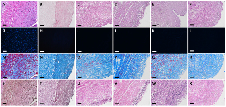
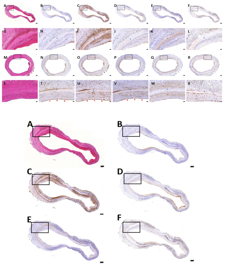
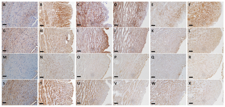
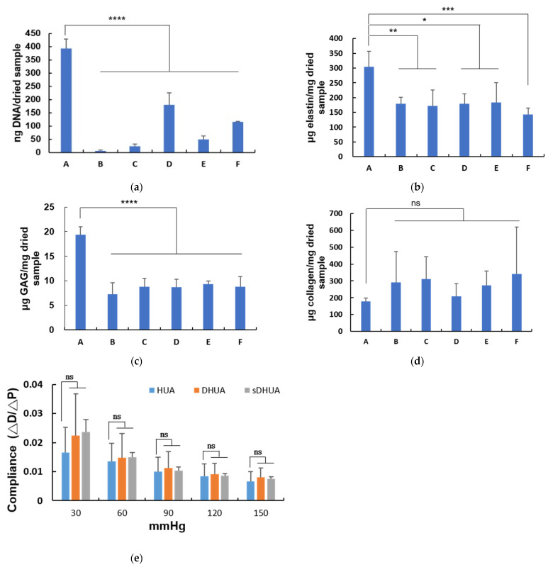
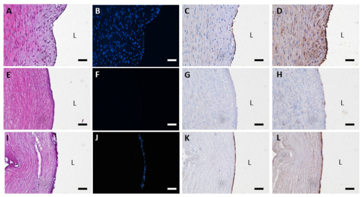
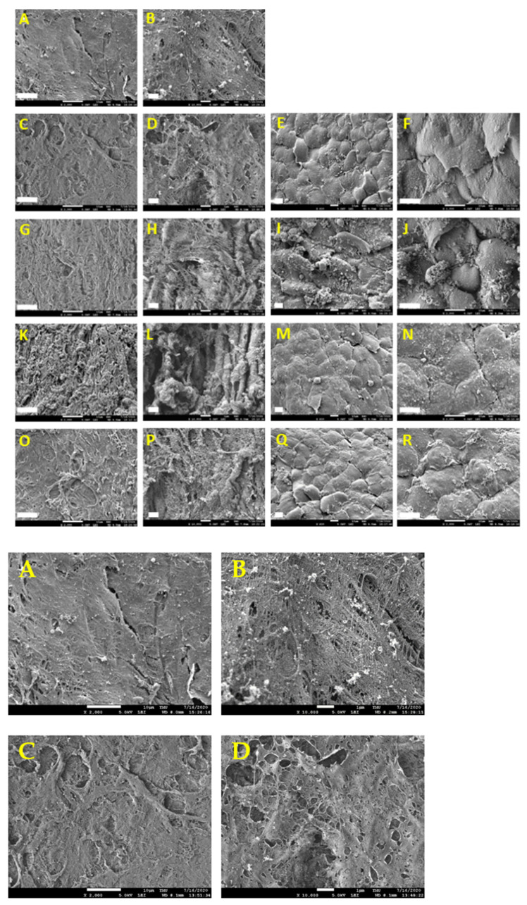
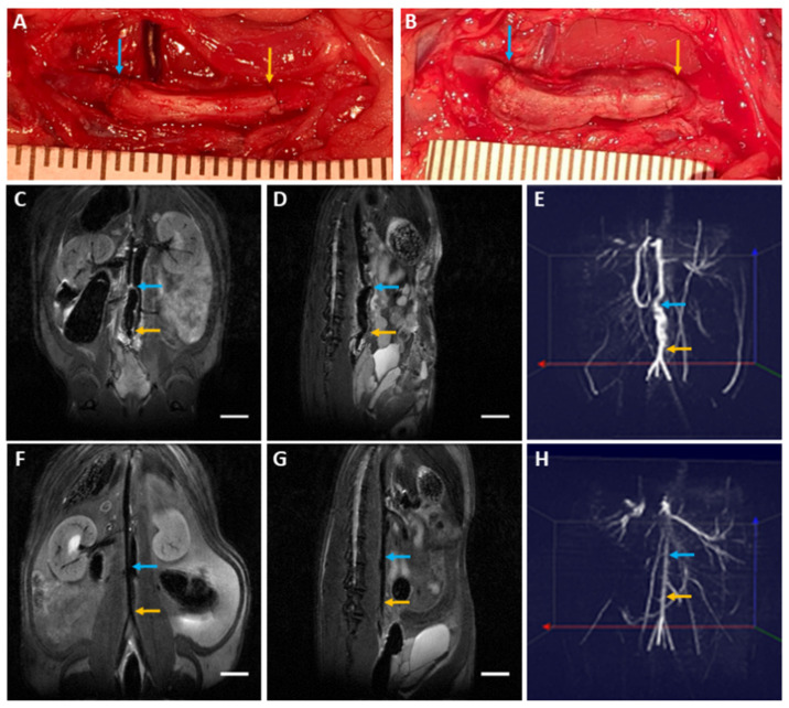
Decellularized vascular grafts are useful for the construction of biological small-diameter tissue-engineered vascular grafts (≤6 mm). Traditional chemical decellularization requires a long treatment time, which may damage the structure and alter the mechanical properties. Decellularization using sonication is expected to solve this problem. The aim of this study was to develop an effective decellularization method using ultrasound followed by washing. Different power values of sonication at 40 kHz were tested for 2, 4, and 8 h followed by a washing procedure. The efficacy of sonication of decellularized human umbilical artery (sDHUA) was evaluated via DNA content, histological staining, mechanical properties, and biocompatibility. The sDHUAs were further implanted into rats for up to 90 days and magnetic resonance angiography (MRA) was performed for the implanted grafts. The results demonstrated that treatment of human umbilical artery (HUA) by sonication at ultrasonic power of 204 W for 4 h followed by washing for 24 h in 2% SDS buffer could eliminate more than 90% of cells and retain similar mechanical properties of the HUA. Recellularization was assessed by scanning electron microscopy (SEM), which indicated that sDHUA provided niches for human umbilical vein endothelial cells (HUVECs) to reside, indicating in vitro cytocompatibility. Further implantation tests also indicated the fitness of the sonication-treated HUA as a scaffold for small-caliber tissue engineering vascular grafts.
去细胞血管移植物可用于构建生物小口径组织工程血管移植物(≤6毫米)。传统的化学去细胞方法需要较长的处理时间,这可能会破坏结构并改变机械性能。使用超声进行去细胞有望解决这个问题。本研究的目的是开发一种有效的超声去细胞方法,随后进行冲洗。测试了40kHz超声不同功率值处理2、4和8小时,然后进行冲洗程序。通过DNA含量、组织学染色、机械性能和生物相容性评估去细胞人脐动脉(sDHUA)的超声处理效果。将sDHUA进一步植入大鼠体内长达90天,并对植入的移植物进行磁共振血管造影(MRA)。结果表明,以204W超声功率对人脐动脉(HUA)进行4小时超声处理,然后在2%SDS缓冲液中冲洗24小时,可以去除90%以上的细胞,并保留HUA相似的机械性能。通过扫描电子显微镜(SEM)评估再细胞化,结果表明sDHUA为人类脐静脉内皮细胞(HUVECs)提供了驻留的微环境,表明其具有体外细胞相容性。进一步的植入测试也表明,超声处理后的HUA适合作为小口径组织工程血管移植物的支架。