• 文献检索
  • 文档翻译
  • 深度研究
  • 学术资讯
  • Suppr Zotero 插件Zotero 插件
  • 邀请有礼
  • 套餐&价格
  • 历史记录
应用&插件
Suppr Zotero 插件Zotero 插件浏览器插件Mac 客户端Windows 客户端微信小程序
定价
高级版会员购买积分包购买API积分包
服务
文献检索文档翻译深度研究API 文档MCP 服务
关于我们
关于 Suppr公司介绍联系我们用户协议隐私条款
关注我们

Suppr 超能文献

核心技术专利:CN118964589B侵权必究
粤ICP备2023148730 号-1Suppr @ 2026

文献检索

告别复杂PubMed语法,用中文像聊天一样搜索,搜遍4000万医学文献。AI智能推荐,让科研检索更轻松。

立即免费搜索

文件翻译

保留排版,准确专业,支持PDF/Word/PPT等文件格式,支持 12+语言互译。

免费翻译文档

深度研究

AI帮你快速写综述,25分钟生成高质量综述,智能提取关键信息,辅助科研写作。

立即免费体验

光学相干断层扫描衍生的斑块形态与血流储备分数测定的中度冠状动脉狭窄的血流动力学相关性

OCT-Derived Plaque Morphology and FFR-Determined Hemodynamic Relevance in Intermediate Coronary Stenoses.

作者信息

Tomaniak Mariusz, Ochijewicz Dorota, Kołtowski Łukasz, Rdzanek Adam, Pietrasik Arkadiusz, Jąkała Jacek, Slezak Magdalena, Malinowski Krzysztof P, Zaleska Martyna, Maksym Jakub, Barus Piotr, Roleder Tomasz, Filipiak Krzysztof J, Opolski Grzegorz, Kochman Janusz

机构信息

First Department of Cardiology, Medical University of Warsaw, 02-097 Warsaw, Poland.

Krakow Cardiovascular Research Institute, 30-055 Krakow, Poland.

出版信息

J Clin Med. 2021 May 28;10(11):2379. doi: 10.3390/jcm10112379.

DOI:10.3390/jcm10112379
PMID:34071299
原文链接:https://pmc.ncbi.nlm.nih.gov/articles/PMC8197966/
Abstract

BACKGROUND

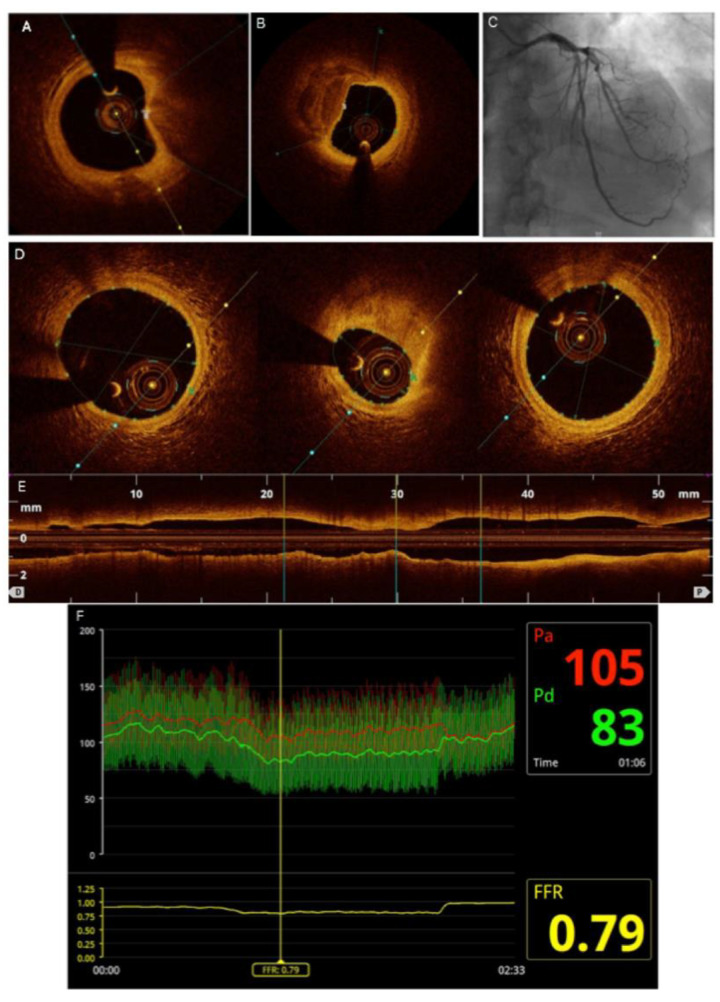
optical coherence tomography (OCT) might allow identifying lesion features reportedly associated with plaque vulnerability and increased risk of clinical events. Previous studies on correlation between OCT and functional lesion significance indices reported contradictory results, yet integration of complementary information from both modalities is gaining increased interest. The aim of the study was to compare plaque morphology using OCT in hemodynamically relevant vs. non-relevant lesions by fractional flow reserve (FFR).

METHODS

consecutive patients with intermediate grade coronary stenoses by angiography were evaluated by both FFR and OCT in this single-center study. Stenoses were labeled hemodynamically relevant in case of the FFR ≤ 0.80. Minimal lumen area (MLA), fibrous cap thickness (FCT), minimal cap thickness over the calcium, angle of the calcium, and necrotic core within the lesions were evaluated.

RESULTS

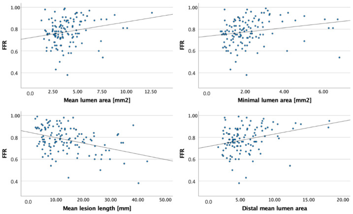
a total of 105 patients (124 vessels) were analyzed. Of them, 65 patients were identified with at least one lesion identified as hemodynamically relevant by FFR (72 vessels, 58.1%). Lesions with FFR ≤0.80 presented with lower mean and minimal lumen area (3.46 ± 1.29 vs. 4.65 ± 2.19, =0.001 and 1.84 ± 0.97 vs. 2.66 ± 1.40, = 0.001) compared to patients with FFR > 0.80. No differences were found between groups in the mean and minimal FCT, mean, and maximal necrotic core, calcium angle, as well as the overall rate of calcified and lipid plaques.

CONCLUSION

hemodynamic relevance of intermediate grade lesions correlated moderately with the luminal assessment by OCT. No differences were identified in the plaque morphology between relevant and non-relevant coronary stenoses by FFR.

摘要

背景

光学相干断层扫描(OCT)或许能够识别出据报道与斑块易损性及临床事件风险增加相关的病变特征。先前关于OCT与功能性病变严重程度指标之间相关性的研究报告了相互矛盾的结果,然而,整合来自这两种模式的互补信息正日益受到关注。本研究的目的是通过血流储备分数(FFR),使用OCT比较血流动力学相关病变与非相关病变的斑块形态。

方法

在这项单中心研究中,对通过血管造影显示为中度冠状动脉狭窄的连续患者进行了FFR和OCT评估。若FFR≤0.80,则将狭窄标记为血流动力学相关。评估了病变内的最小管腔面积(MLA)、纤维帽厚度(FCT)、钙化上方的最小帽厚度、钙化角度和坏死核心。

结果

共分析了105例患者(124支血管)。其中,65例患者被识别出至少有一个病变被FFR确定为血流动力学相关(72支血管,58.1%)。与FFR>0.80的患者相比,FFR≤0.80的病变平均管腔面积和最小管腔面积更低(分别为3.46±1.29 vs. 4.65±2.19,P = 0.001;1.84±0.97 vs. 2.66±1.40,P = 0.001)。两组在平均和最小FCT、平均和最大坏死核心、钙化角度以及钙化和脂质斑块的总体发生率方面未发现差异。

结论

中度病变的血流动力学相关性与OCT的管腔评估中度相关。通过FFR,相关和非相关冠状动脉狭窄之间的斑块形态未发现差异。

https://cdn.ncbi.nlm.nih.gov/pmc/blobs/2ffc/8197966/16af292d74e6/jcm-10-02379-g003.jpg
https://cdn.ncbi.nlm.nih.gov/pmc/blobs/2ffc/8197966/4aefcb10f8b7/jcm-10-02379-g001.jpg
https://cdn.ncbi.nlm.nih.gov/pmc/blobs/2ffc/8197966/ab3cc70a25df/jcm-10-02379-g002.jpg
https://cdn.ncbi.nlm.nih.gov/pmc/blobs/2ffc/8197966/16af292d74e6/jcm-10-02379-g003.jpg
https://cdn.ncbi.nlm.nih.gov/pmc/blobs/2ffc/8197966/4aefcb10f8b7/jcm-10-02379-g001.jpg
https://cdn.ncbi.nlm.nih.gov/pmc/blobs/2ffc/8197966/ab3cc70a25df/jcm-10-02379-g002.jpg
https://cdn.ncbi.nlm.nih.gov/pmc/blobs/2ffc/8197966/16af292d74e6/jcm-10-02379-g003.jpg

相似文献

1
OCT-Derived Plaque Morphology and FFR-Determined Hemodynamic Relevance in Intermediate Coronary Stenoses.光学相干断层扫描衍生的斑块形态与血流储备分数测定的中度冠状动脉狭窄的血流动力学相关性
J Clin Med. 2021 May 28;10(11):2379. doi: 10.3390/jcm10112379.
2
Relationship between optical coherence tomography derived intraluminal and intramural criteria and haemodynamic relevance as determined by fractional flow reserve in intermediate coronary stenoses of patients with type 2 diabetes.光学相干断层成像技术(OCT)测量的管腔和管壁标准与血流储备分数(FFR)在 2 型糖尿病患者中等程度冠状动脉狭窄中的相关性。
Heart. 2013 May;99(10):700-7. doi: 10.1136/heartjnl-2013-303616. Epub 2013 Mar 30.
3
Multimodality OCT, IVUS and FFR evaluation of coronary intermediate grade lesions in women vs. men.多模态光学相干断层扫描(OCT)、血管内超声(IVUS)和血流储备分数(FFR)对女性与男性冠状动脉中度病变的评估
Front Cardiovasc Med. 2023 Jun 22;10:1021023. doi: 10.3389/fcvm.2023.1021023. eCollection 2023.
4
Correlation between fractional flow reserve and instantaneous wave-free ratio with morphometric assessment by optical coherence tomography in diabetic patients.糖尿病患者光学相干断层成像形态测量评估的血流储备分数与瞬时无波比的相关性。
Int J Cardiovasc Imaging. 2020 Jul;36(7):1193-1201. doi: 10.1007/s10554-020-01819-3. Epub 2020 Mar 27.
5
Evaluation of hemodynamically severe coronary stenosis as determined by fractional flow reserve with frequency domain optical coherence tomography measured anatomical parameters.通过频域光学相干断层扫描测量的解剖学参数,对血流动力学严重冠状动脉狭窄进行血流储备分数测定的评估。
J Cardiol. 2014 Jul;64(1):19-24. doi: 10.1016/j.jjcc.2013.11.009. Epub 2013 Dec 22.
6
Multimodality imaging of intermediate lesions: Data from fractional flow reserve, optical coherence tomography, near-infrared spectroscopy-intravascular ultrasound.中等病变的多模态成像:来自血流储备分数、光学相干断层扫描、近红外光谱-血管内超声的数据。
Cardiol J. 2018;25(2):196-202. doi: 10.5603/CJ.a2017.0082. Epub 2017 Jul 17.
7
Relationship between optical coherence tomography-derived morphological criteria and functional relevance as determined by fractional flow reserve.光学相干断层扫描衍生的形态学标准与通过血流储备分数确定的功能相关性之间的关系。
J Cardiol. 2018 Apr;71(4):359-366. doi: 10.1016/j.jjcc.2017.09.010. Epub 2017 Nov 2.
8
Optical coherence tomography enables more accurate detection of functionally significant intermediate non-left main coronary artery stenoses than intravascular ultrasound: A meta-analysis of 6919 patients and 7537 lesions.光学相干断层扫描比血管内超声能更准确地检测出具有功能意义的非左主干冠状动脉中度狭窄:一项对6919例患者和7537处病变的荟萃分析。
Int J Cardiol. 2020 Feb 15;301:226-234. doi: 10.1016/j.ijcard.2019.09.067. Epub 2019 Oct 9.
9
Non-invasive prediction of hemodynamically significant coronary artery stenoses by contrast density difference in coronary CT angiography.通过冠状动脉CT血管造影中的对比剂密度差异对血流动力学显著的冠状动脉狭窄进行无创预测。
Eur J Radiol. 2015 Aug;84(8):1502-1508. doi: 10.1016/j.ejrad.2015.04.024. Epub 2015 May 11.
10
Association between fractional flow reserve and coronary plaque characteristics assessed by optical coherence tomography.光学相干断层扫描评估的血流储备分数与冠状动脉斑块特征之间的关联。
J Cardiol. 2016 Oct;68(4):342-5. doi: 10.1016/j.jjcc.2015.10.012. Epub 2015 Nov 18.

引用本文的文献

1
When Functional Assessment Meets Intravascular Imaging in Patients with Coronary Artery Disease.当功能评估与冠状动脉疾病患者的血管内成像相遇时。
J Cardiovasc Dev Dis. 2025 Aug 20;12(8):319. doi: 10.3390/jcdd12080319.
2
Association between optical coherence tomography and functionally severe stenosis assessed by quantitative flow ratio in coronary intermediate lesions.光学相干断层扫描与通过冠状动脉中度病变的定量血流比评估的功能严重狭窄之间的关联。
PLoS One. 2025 Jun 10;20(6):e0324872. doi: 10.1371/journal.pone.0324872. eCollection 2025.
3
Role of OCT in Assessing Vasa Vasorum in Chronic Coronary Syndrome: Insights from Long-Term Follow-Up.

本文引用的文献

1
Quantitative Flow Ratio Is Related to Intraluminal Coronary Stenosis Parameters as Assessed with Optical Coherence Tomography.定量血流比与光学相干断层扫描评估的冠状动脉管腔内狭窄参数相关。
J Clin Med. 2021 Apr 24;10(9):1856. doi: 10.3390/jcm10091856.
2
Prognostic irrelevance of plaque vulnerability following plaque sealing in high-risk patients with type 2 diabetes: an optical coherence tomography study.斑块易损性在斑块封闭后对 2 型糖尿病高危患者预后无影响:光学相干断层扫描研究。
Cardiovasc Diabetol. 2020 Nov 12;19(1):192. doi: 10.1186/s12933-020-01168-4.
3
Percutaneous Coronary Intervention for Vulnerable Coronary Atherosclerotic Plaque.
光学相干断层扫描在评估慢性冠状动脉综合征中滋养血管的作用:长期随访的见解
J Clin Med. 2025 Feb 26;14(5):1560. doi: 10.3390/jcm14051560.
4
Intracoronary Imaging: Current Practice and Future Perspectives.冠状动脉内成像:当前实践与未来展望。
Rev Cardiovasc Med. 2023 Feb 2;24(2):39. doi: 10.31083/j.rcm2402039. eCollection 2023 Feb.
5
The Plaque Analysis Classifies the Coronary Artery Disease-Reporting and Data System (CAD-RADS) Stenosis and Plaque Burden Categories: Association of the Plaque Features, Fat Attenuation Index, Coronary Computed Tomography Fractional Flow Reserve, and the Combination of Stenosis and Calcification.斑块分析分类冠状动脉疾病报告和数据系统 (CAD-RADS) 狭窄和斑块负荷分类:斑块特征、脂肪衰减指数、冠状动脉计算机断层血流储备分数的关联,以及狭窄和钙化的组合。
Clin Cardiol. 2024 Jun;47(6):e24305. doi: 10.1002/clc.24305.
6
Comparison between Imaging and Physiology in Guiding Coronary Revascularization: A Meta-Analysis.影像学与生理学在指导冠状动脉血运重建中的比较:一项荟萃分析。
J Clin Med. 2024 Apr 24;13(9):2504. doi: 10.3390/jcm13092504.
7
Renal Dysfunction Increases Risk of Adverse Cardiovascular Events in 5-Year Follow-Up Study of Intermediate Coronary Artery Lesions.肾功能障碍增加了 5 年随访研究中中等冠状动脉病变患者不良心血管事件的风险。
Med Sci Monit. 2024 May 9;30:e943956. doi: 10.12659/MSM.943956.
8
Association between vessel-specific coronary Aggregated plaque burden, Agatston score and hemodynamic significance of coronary disease (The CAPTivAte study).血管特异性冠状动脉总斑块负荷、阿加斯顿评分与冠心病血流动力学意义之间的关联(CAPTivAte研究)
Int J Cardiol Heart Vasc. 2024 Mar 10;51:101384. doi: 10.1016/j.ijcha.2024.101384. eCollection 2024 Apr.
9
Assessment of the functional severity of coronary lesions from optical coherence tomography based on ensembled learning.基于集成学习的光学相干断层成像术评估冠状动脉病变的功能严重程度。
Biomed Eng Online. 2023 Dec 16;22(1):127. doi: 10.1186/s12938-023-01192-x.
10
Multimodality OCT, IVUS and FFR evaluation of coronary intermediate grade lesions in women vs. men.多模态光学相干断层扫描(OCT)、血管内超声(IVUS)和血流储备分数(FFR)对女性与男性冠状动脉中度病变的评估
Front Cardiovasc Med. 2023 Jun 22;10:1021023. doi: 10.3389/fcvm.2023.1021023. eCollection 2023.
经皮冠状动脉介入治疗易损冠状动脉粥样硬化斑块
J Am Coll Cardiol. 2020 Nov 17;76(20):2289-2301. doi: 10.1016/j.jacc.2020.09.547. Epub 2020 Oct 15.
4
Vulnerable plaques and patients: state-of-the-art.易损斑块与患者:现状分析。
Eur Heart J. 2020 Aug 14;41(31):2997-3004. doi: 10.1093/eurheartj/ehaa227.
5
Correlation between fractional flow reserve and instantaneous wave-free ratio with morphometric assessment by optical coherence tomography in diabetic patients.糖尿病患者光学相干断层成像形态测量评估的血流储备分数与瞬时无波比的相关性。
Int J Cardiovasc Imaging. 2020 Jul;36(7):1193-1201. doi: 10.1007/s10554-020-01819-3. Epub 2020 Mar 27.
6
Relationship between coronary plaque morphology of the left anterior descending artery and 12 months clinical outcome: the CLIMA study.左前降支冠状动脉斑块形态与 12 个月临床结局的关系:CLIMA 研究。
Eur Heart J. 2020 Jan 14;41(3):383-391. doi: 10.1093/eurheartj/ehz520.
7
Diagnostic accuracy of intracoronary optical coherence tomography-derived fractional flow reserve for assessment of coronary stenosis severity.冠状动脉光学相干断层成像衍生的血流储备分数评估冠状动脉狭窄严重程度的诊断准确性。
EuroIntervention. 2019 Jun 20;15(2):189-197. doi: 10.4244/EIJ-D-19-00182.
8
Comparison between instantaneous wave-free ratio versus morphometric assessments by intracoronary imaging.冠状动脉内成像评估瞬时无波比值与形态学指标的比较。
Heart Vessels. 2019 Jun;34(6):926-935. doi: 10.1007/s00380-018-1320-4. Epub 2018 Dec 7.
9
2018 ESC/EACTS Guidelines on myocardial revascularization.2018年欧洲心脏病学会/欧洲心胸外科学会心肌血运重建指南。
Eur Heart J. 2019 Jan 7;40(2):87-165. doi: 10.1093/eurheartj/ehy394.
10
Clinical use of intracoronary imaging. Part 1: guidance and optimization of coronary interventions. An expert consensus document of the European Association of Percutaneous Cardiovascular Interventions.冠状动脉内影像学的临床应用。第 1 部分:冠状动脉介入治疗的指导和优化。欧洲经皮心血管介入治疗协会的专家共识文件。
EuroIntervention. 2018 Aug 20;14(6):656-677. doi: 10.4244/EIJY18M06_01.