David H. Koch Institute for Integrative Cancer Research, Massachusetts Institute of Technology, Cambridge, MA 02142.
Department of Biology, Massachusetts Institute of Technology, Cambridge, MA 02142.
Proc Natl Acad Sci U S A. 2021 Jun 8;118(23). doi: 10.1073/pnas.2019740118.
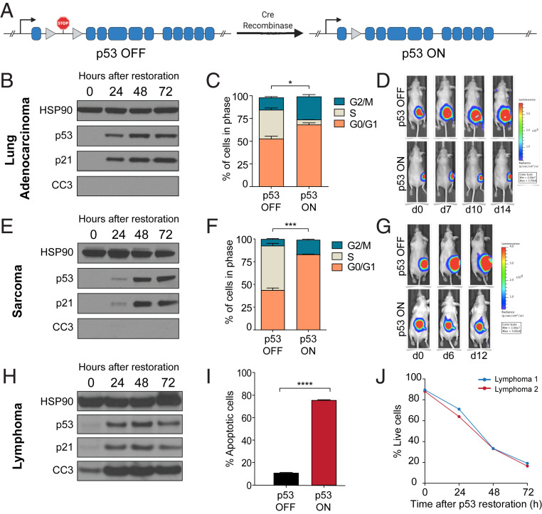
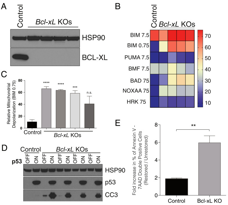
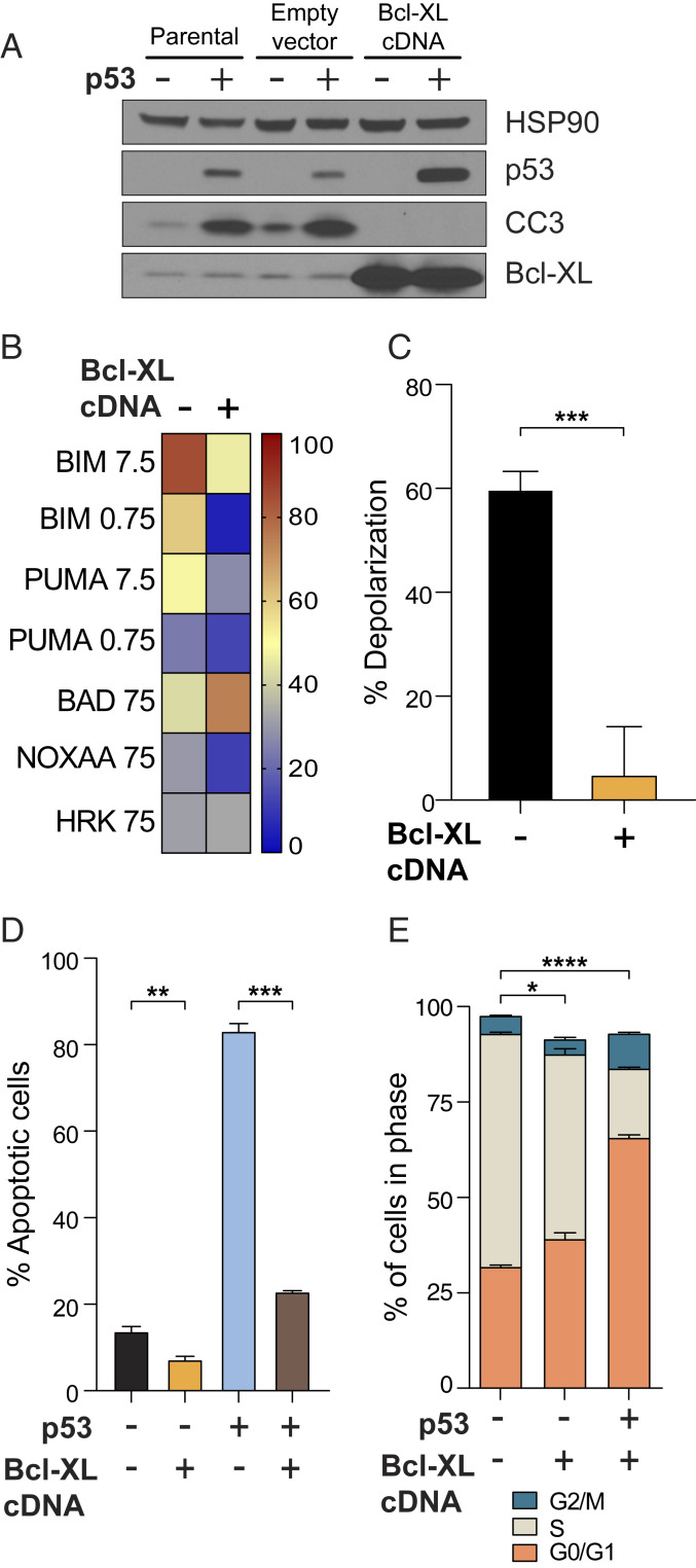
Reactivation of p53 in established tumors typically results in one of two cell fates, cell cycle arrest or apoptosis, but it remains unclear how this cell fate is determined. We hypothesized that high mitochondrial priming prior to p53 reactivation would lead to apoptosis, while low priming would lead to survival and cell cycle arrest. Using a panel of Kras-driven, p53 restorable cell lines derived from genetically engineered mouse models of lung adenocarcinoma and sarcoma (both of which undergo cell cycle arrest upon p53 restoration), as well as lymphoma (which instead undergo apoptosis), we show that the level of mitochondrial apoptotic priming is a critical determinant of p53 reactivation outcome. Cells with high initial priming (e.g., lymphomas) lacked sufficient reserve antiapoptotic capacity and underwent apoptosis after p53 restoration. Forced BCL-2 or BCL-XL expression reduced priming and resulted in survival and cell cycle arrest. Cells with low initial priming (e.g., lung adenocarcinoma and sarcoma) survived and proceeded to arrest in the cell cycle. When primed by inhibition of their antiapoptotic proteins using genetic (BCL-2 or BCL-XL deletion or BAD overexpression) or pharmacologic (navitoclax) means, apoptosis resulted upon p53 restoration in vitro and in vivo. These data demonstrate that mitochondrial apoptotic priming is a key determining factor of cell fate upon p53 activation. Moreover, it is possible to enforce apoptotic cell fate following p53 activation in less primed cells using p53-independent drugs that increase apoptotic priming, including BH3 mimetic drugs.
已建立的肿瘤中 p53 的再激活通常导致两种细胞命运之一,即细胞周期停滞或细胞凋亡,但尚不清楚这种细胞命运是如何决定的。我们假设在 p53 再激活之前,高线粒体凋亡启动会导致细胞凋亡,而低启动则会导致细胞存活和细胞周期停滞。我们使用一组由遗传工程小鼠模型中的肺腺癌和肉瘤(两者在 p53 恢复时都经历细胞周期停滞)以及淋巴瘤(相反经历细胞凋亡)衍生的 Kras 驱动、p53 可恢复的细胞系进行了研究,表明线粒体凋亡启动的水平是 p53 再激活结果的关键决定因素。初始启动水平较高的细胞(例如淋巴瘤)缺乏足够的储备抗凋亡能力,并在 p53 恢复后发生细胞凋亡。强制表达 BCL-2 或 BCL-XL 会降低启动水平,并导致细胞存活和细胞周期停滞。初始启动水平较低的细胞(例如肺腺癌和肉瘤)存活下来,并继续在细胞周期中停滞。当通过遗传(BCL-2 或 BCL-XL 缺失或 BAD 过表达)或药理学(navitoclax)手段抑制其抗凋亡蛋白来启动时,p53 在体外和体内恢复后会导致细胞凋亡。这些数据表明,线粒体凋亡启动是 p53 激活时细胞命运的关键决定因素。此外,使用可增加凋亡启动的 p53 非依赖性药物(包括 BH3 模拟药物),可以在启动水平较低的细胞中强制实施 p53 激活后的凋亡细胞命运。