Strange Daniel P, Jiyarom Boonyanudh, Sadri-Ardekani Hooman, Cazares Lisa H, Kenny Tara A, Ward Michael D, Verma Saguna
Department of Tropical Medicine, Medical Microbiology, and Pharmacology, John A. Burns School of Medicine, University of Hawai'i at Mãnoa, Honolulu, HI, United States.
Wake Forest Institute for Regenerative Medicine, Wake Forest School of Medicine, Winston-Salem, NC, United States.
Front Microbiol. 2021 May 17;12:667146. doi: 10.3389/fmicb.2021.667146. eCollection 2021.
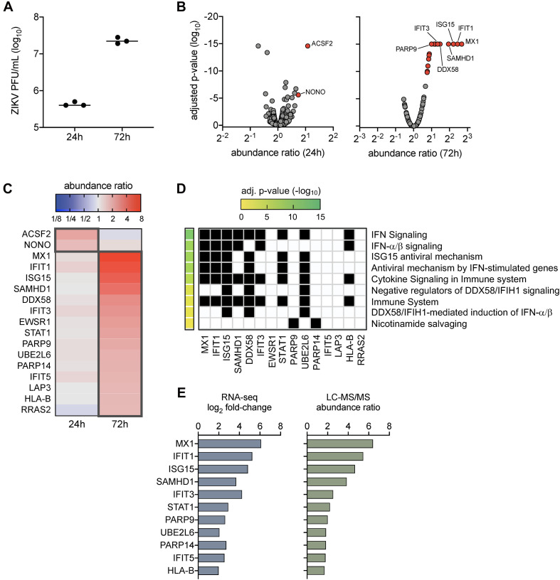
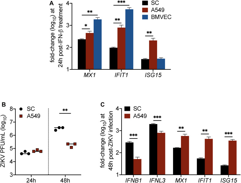
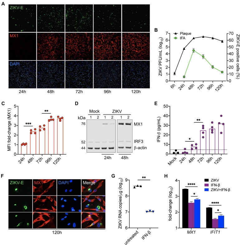
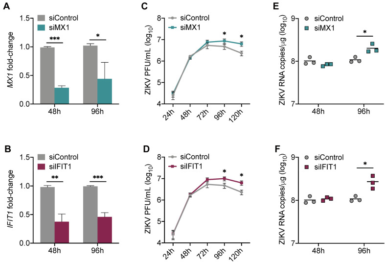
Zika virus (ZIKV) is unique among mosquito-borne flaviviruses in its ability to be sexually transmitted. The testes have been implicated as sites of long-term ZIKV replication, and our previous studies have identified Sertoli cells (SC), the nurse cells of the seminiferous epithelium that govern spermatogenesis, as major targets of ZIKV infection. To improve our understanding of the interaction of ZIKV with human SC, we analyzed ZIKV-induced proteome changes in these cells using high-throughput liquid chromatography-tandem mass spectrometry (LC-MS/MS). Our data demonstrated that interferon (IFN) signaling was the most significantly enriched pathway and the antiviral proteins MX1 and IFIT1 were among the top upregulated proteins in SC following ZIKV infection. The dynamic between IFN response and ZIKV infection kinetics in SC remains unclear, therefore we further determined whether MX1 and IFIT1 serve as antiviral effectors against ZIKV. We found that increased levels of MX1 at the later time points of infection coincided with diminished ZIKV infection while the silencing of and enhanced peak ZIKV propagation in SC. Furthermore, although IFN-I exposure was found to significantly hinder ZIKV replication in SC, IFN response was attenuated in these cells as compared to other cell types. The data in this study highlight IFN-I as a driver of the antiviral state that limits ZIKV infection in SC and suggests that MX1 and IFIT1 function as antiviral effectors against ZIKV in SC. Collectively, this study provides important biological insights into the response of SC to ZIKV infection and the ability of the virus to persist in the testes.
寨卡病毒(ZIKV)在蚊媒传播的黄病毒中独具能够通过性传播的特性。睾丸被认为是寨卡病毒长期复制的场所,我们之前的研究已确定支持细胞(SC),即生精上皮中控制精子发生的滋养细胞,是寨卡病毒感染的主要靶标。为了增进我们对寨卡病毒与人类支持细胞相互作用的理解,我们使用高通量液相色谱 - 串联质谱法(LC-MS/MS)分析了寨卡病毒诱导的这些细胞中的蛋白质组变化。我们的数据表明,干扰素(IFN)信号通路是最显著富集的通路,抗病毒蛋白MX1和IFIT1是寨卡病毒感染后支持细胞中上调最明显的蛋白质之一。支持细胞中IFN反应与寨卡病毒感染动力学之间的动态关系仍不清楚,因此我们进一步确定MX1和IFIT1是否作为针对寨卡病毒的抗病毒效应因子。我们发现,在感染后期MX1水平升高与寨卡病毒感染减少同时出现,而MX1和IFIT1的沉默增强了支持细胞中寨卡病毒的峰值传播。此外,虽然发现I型干扰素暴露显著阻碍支持细胞中的寨卡病毒复制,但与其他细胞类型相比,这些细胞中的IFN反应减弱。本研究中的数据突出了I型干扰素作为限制支持细胞中寨卡病毒感染的抗病毒状态驱动因子的作用,并表明MX1和IFIT1在支持细胞中作为针对寨卡病毒的抗病毒效应因子发挥作用。总体而言,本研究为支持细胞对寨卡病毒感染的反应以及病毒在睾丸中持续存在的能力提供了重要的生物学见解。