EPSRC Centre for Doctoral Training in Intelligent, Integrated Imaging in Healthcare (i4health), University College London, Gower Street, London WC1E 6BT, United Kingdom; Functional Neurosurgery Unit, Department of Clinical and Motor Neurosciences, UCL Institute of Neurology, Queen Square, WC1N 3BG London, United Kingdom; Wellcome Centre for Human Neuroimaging, 12 Queen Square, London WC1N 3AR, United Kingdom.
Functional Neurosurgery Unit, Department of Clinical and Motor Neurosciences, UCL Institute of Neurology, Queen Square, WC1N 3BG London, United Kingdom.
Neuroimage. 2021 Sep;238:118231. doi: 10.1016/j.neuroimage.2021.118231. Epub 2021 Jun 2.
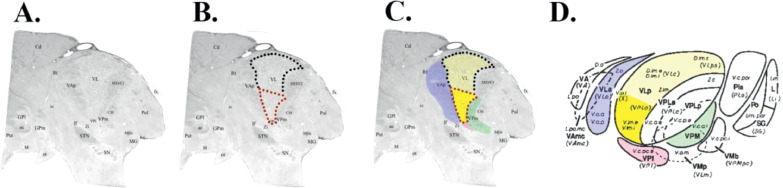
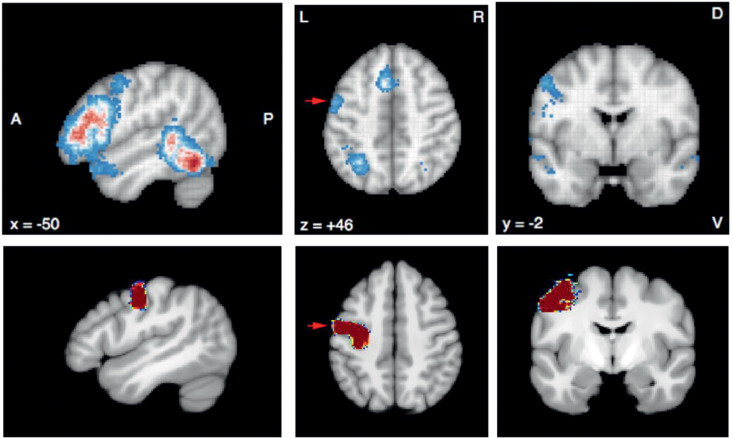
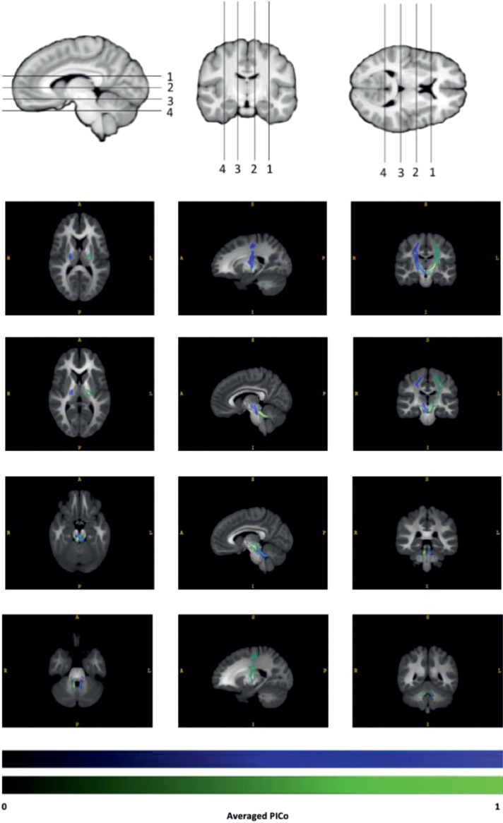
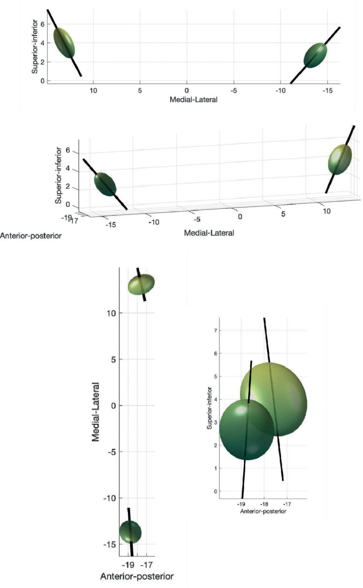
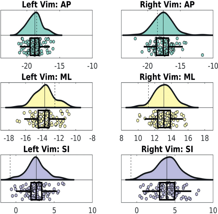
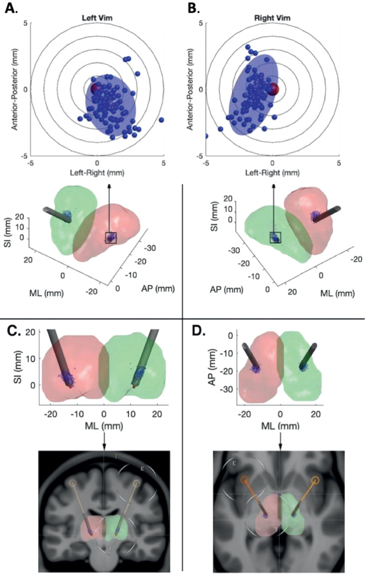
The ventralis intermedius nucleus (Vim) is centrally placed in the dentato-thalamo-cortical pathway (DTCp) and is a key surgical target in the treatment of severe medically refractory tremor. It is not visible on conventional MRI sequences; consequently, stereotactic targeting currently relies on atlas-based coordinates. This fails to capture individual anatomical variability, which may lead to poor long-term clinical efficacy. Probabilistic tractography, combined with known anatomical connectivity, enables localisation of thalamic nuclei at an individual subject level. There are, however, a number of confounds associated with this technique that may influence results. Here we focused on an established method, using probabilistic tractography to reconstruct the DTCp, to identify the connectivity-defined Vim (cd-Vim) in vivo. Using 100 healthy individuals from the Human Connectome Project, our aim was to quantify cd-Vim variability across this population, measure the discrepancy with atlas-defined Vim (ad-Vim), and assess the influence of potential methodological confounds. We found no significant effect of any of the confounds. The mean cd-Vim coordinate was located within 1.88 mm (left) and 2.12 mm (right) of the average midpoint and 3.98 mm (left) and 5.41 mm (right) from the ad-Vim coordinates. cd-Vim location was more variable on the right, which reflects hemispheric asymmetries in the probabilistic DTC reconstructed. The method was reproducible, with no significant cd-Vim location differences in a separate test-retest cohort. The superior cerebellar peduncle was identified as a potential source of artificial variance. This work demonstrates significant individual anatomical variability of the cd-Vim that atlas-based coordinate targeting fails to capture. This variability was not related to any methodological confound tested. Lateralisation of cerebellar functions, such as speech, may contribute to the observed asymmetry. Tractography-based methods seem sensitive to individual anatomical variability that is missed by conventional neurosurgical targeting; these findings may form the basis for translational tools to improve efficacy and reduce side-effects of thalamic surgery for tremor.
腹中间核(Vim)位于齿状核-丘脑-皮质通路(DTCp)的中心,是治疗严重药物难治性震颤的关键手术靶点。它在常规 MRI 序列中不可见;因此,目前的立体定向靶向依赖于基于图谱的坐标。这未能捕捉到个体解剖结构的可变性,这可能导致长期临床疗效不佳。概率追踪技术,结合已知的解剖连接性,可以在个体水平上定位丘脑核。然而,该技术存在一些与结果相关的混淆因素。在这里,我们专注于一种已建立的方法,使用概率追踪来重建 DTCp,以在体内识别连接定义的 Vim(cd-Vim)。使用来自人类连接组计划的 100 名健康个体,我们的目标是量化该人群中 cd-Vim 的可变性,测量与图谱定义的 Vim(ad-Vim)的差异,并评估潜在方法学混淆因素的影响。我们发现任何混淆因素都没有显著影响。cd-Vim 坐标的平均值位于平均中点的 1.88mm(左侧)和 2.12mm(右侧)内,与 ad-Vim 坐标的距离为 3.98mm(左侧)和 5.41mm(右侧)。cd-Vim 的位置在右侧更具可变性,这反映了概率 DTC 重建中的半球不对称性。该方法具有可重复性,在另一个测试-重测队列中,cd-Vim 的位置没有显著差异。上小脑脚被确定为人工方差的潜在来源。这项工作表明,cd-Vim 的个体解剖结构具有显著的可变性,而基于图谱的坐标靶向无法捕捉到这一点。这种可变性与任何测试的方法学混淆因素无关。言语等小脑功能的偏侧化可能导致观察到的不对称性。基于轨迹的方法似乎对常规神经外科靶向方法错过的个体解剖结构可变性敏感;这些发现可能为改善丘脑手术治疗震颤的疗效和减少副作用的转化工具奠定基础。