Viral Immunology Center, Department of Biology, Georgia State University, Atlanta, 30303, Georgia; Department of Ophthalmology, Emory University School of Medicine, Atlanta, 30322, Georgia.
Viral Immunology Center, Department of Biology, Georgia State University, Atlanta, 30303, Georgia.
Exp Eye Res. 2021 Aug;209:108651. doi: 10.1016/j.exer.2021.108651. Epub 2021 Jun 5.
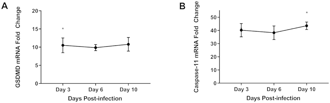
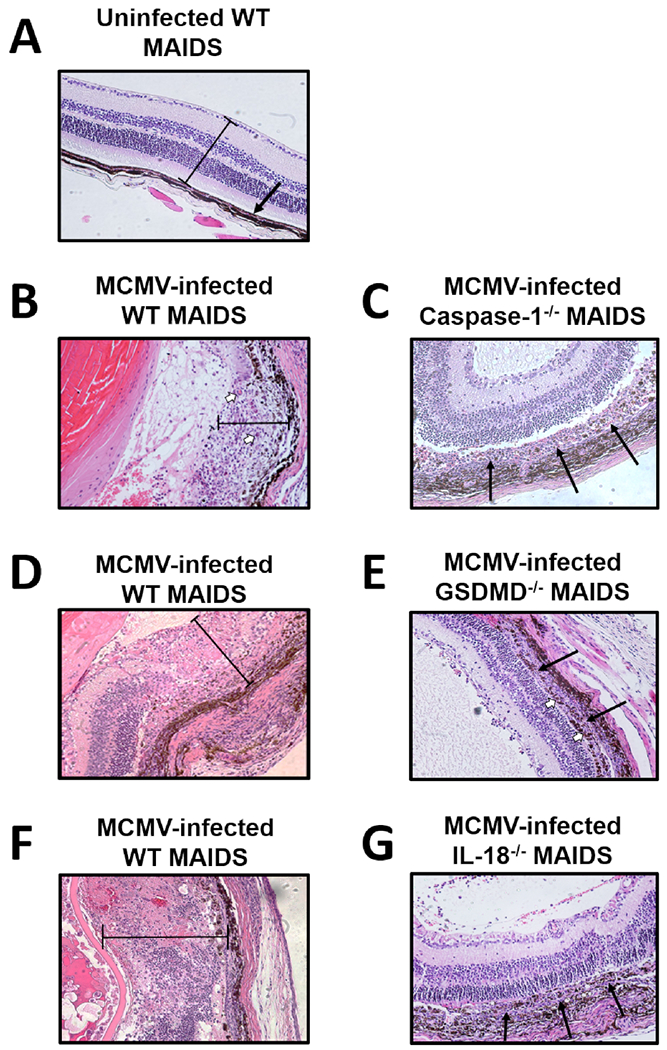
Pyroptosis is a caspase-dependent programmed cell death pathway that initiates and sustains inflammation through release of pro-inflammatory cytokines interleukin (IL)-1β and IL-18 following formation of gasdermin D (GSDMD)-mediated membrane pores. To determine the possible pathogenic contributions of pyroptosis toward development of full-thickness retinal necrosis during AIDS-related human cytomegalovirus retinitis, we performed a series of studies using an established model of experimental murine cytomegalovirus (MCMV) retinitis in mice with retrovirus-induced immunosuppression (MAIDS). Initial investigations demonstrated significant transcription and translation of key pyroptosis-associated genes within the ocular compartments of MCMV-infected eyes of mice with MAIDS. Subsequent investigations compared MCMV-infected eyes of groups of wildtype MAIDS mice with MCMV-infected eyes of groups of caspase-1 MAIDS mice, GSDMD MAIDS mice, or IL-18 MAIDS mice to explore a possible contribution of pyroptosis towards the pathogenesis of MAIDS-related MCMV retinitis. Histopathologic analysis revealed typical full-thickness retinal necrosis in 100% of MCMV-infected eyes of wildtype MAIDS mice. In sharp contrast, none (0%) of MCMV-infected eyes of MAIDS mice that were deficient in either caspase-1, GSDMD, or IL-18 developed full-thickness retinal necrosis but instead exhibited an atypical pattern of retinal disease characterized by thickening and proliferation of the retinal pigmented epithelium layer with relative sparing of the neurosensory retina. Surprisingly, MCMV-infected eyes of all groups of deficient MAIDS mice harbored equivalent intraocular amounts of infectious virus as seen in MCMV-infected eyes of groups of wildtype MAIDS mice despite failure to develop full-thickness retinal necrosis. We conclude that pyroptosis plays a significant role in the development of full-thickness retinal necrosis during the pathogenesis of MAIDS-related MCMV retinitis. This observation may extend to the pathogenesis of AIDS-related HCMV retinitis and other AIDS-related opportunistic virus infections.
细胞焦亡是一种依赖半胱天冬酶的程序性细胞死亡途径,通过形成 Gasdermin D(GSDMD)介导的膜孔后释放促炎细胞因子白细胞介素(IL)-1β和 IL-18,从而引发和维持炎症。为了确定细胞焦亡在艾滋病相关人巨细胞病毒视网膜炎中全层视网膜坏死的发病机制中的可能致病作用,我们使用逆转录病毒诱导免疫抑制(MAIDS)的小鼠实验性巨细胞病毒(MCMV)视网膜炎的既定模型进行了一系列研究。初步研究表明,在 MAIDS 小鼠的感染眼眼部组织中,与细胞焦亡相关的关键基因的转录和翻译均显著增加。随后的研究比较了 MAIDS 野生型小鼠的 MCMV 感染眼与 MAIDS 野生型和 caspase-1 缺陷型、GSDMD 缺陷型或 IL-18 缺陷型 MCMV 感染眼,以探讨细胞焦亡在 MAIDS 相关 MCMV 视网膜炎发病机制中的可能作用。组织病理学分析显示,100%的 MAIDS 野生型 MCMV 感染眼出现典型的全层视网膜坏死。与此形成鲜明对比的是,缺乏 caspase-1、GSDMD 或 IL-18 的 MAIDS 小鼠的 MCMV 感染眼无一例发生全层视网膜坏死,而是表现出一种非典型的视网膜疾病模式,其特征为视网膜色素上皮层增厚和增生,而神经感觉视网膜相对保留。令人惊讶的是,所有缺乏 MAIDS 小鼠的 MCMV 感染眼的眼内病毒载量与 MAIDS 野生型 MCMV 感染眼相当,尽管它们没有发生全层视网膜坏死。我们的结论是,细胞焦亡在 MAIDS 相关 MCMV 视网膜炎发病机制中的全层视网膜坏死的发展中起重要作用。这一观察结果可能扩展到 AIDS 相关 HCMV 视网膜炎和其他 AIDS 相关机会性病毒感染的发病机制。