• 文献检索
  • 文档翻译
  • 深度研究
  • 学术资讯
  • Suppr Zotero 插件Zotero 插件
  • 邀请有礼
  • 套餐&价格
  • 历史记录
应用&插件
Suppr Zotero 插件Zotero 插件浏览器插件Mac 客户端Windows 客户端微信小程序
定价
高级版会员购买积分包购买API积分包
服务
文献检索文档翻译深度研究API 文档MCP 服务
关于我们
关于 Suppr公司介绍联系我们用户协议隐私条款
关注我们

Suppr 超能文献

核心技术专利:CN118964589B侵权必究
粤ICP备2023148730 号-1Suppr @ 2026

文献检索

告别复杂PubMed语法,用中文像聊天一样搜索,搜遍4000万医学文献。AI智能推荐,让科研检索更轻松。

立即免费搜索

文件翻译

保留排版,准确专业,支持PDF/Word/PPT等文件格式,支持 12+语言互译。

免费翻译文档

深度研究

AI帮你快速写综述,25分钟生成高质量综述,智能提取关键信息,辅助科研写作。

立即免费体验

BAFF 信号通过依赖 Pin1 的方式驱动小鼠肾小管上皮细胞的间质转化。

BAFF signaling drives interstitial transformation of mouse renal tubular epithelial cells in a Pin1-dependent manner.

机构信息

Urology Department, Third Affiliated Hospital of Soochow University, 185# Juqian Street, Changzhou, 213003, China.

出版信息

In Vitro Cell Dev Biol Anim. 2021 Jun;57(6):649-659. doi: 10.1007/s11626-021-00598-y. Epub 2021 Jun 14.

DOI:10.1007/s11626-021-00598-y
PMID:34128158
原文链接:https://pmc.ncbi.nlm.nih.gov/articles/PMC8316171/
Abstract

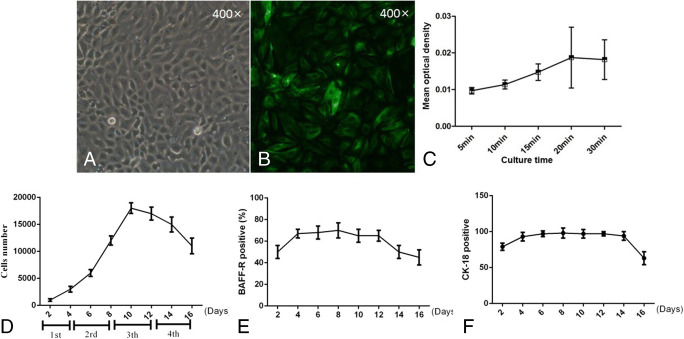
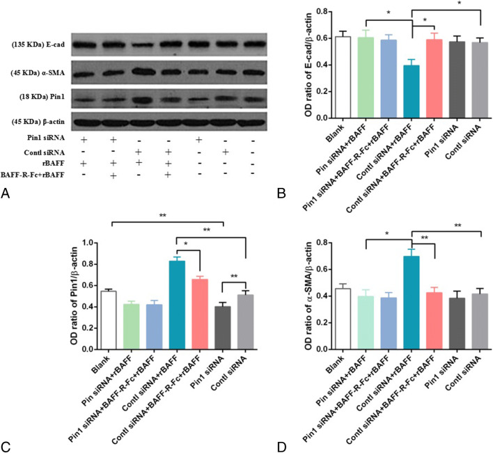
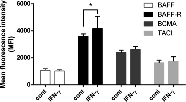
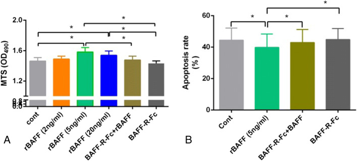
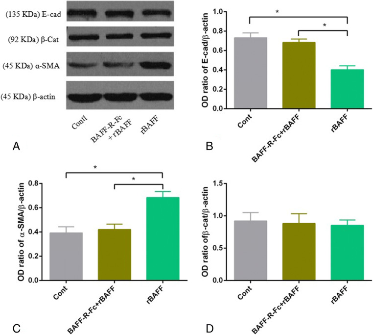
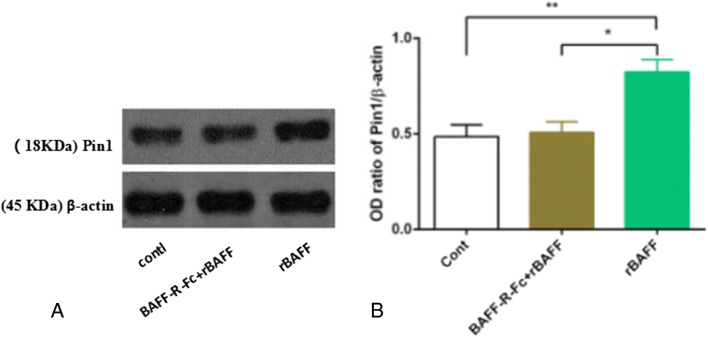
Aberrant expression of B cell-activating factor belonging to TNF superfamily (BAFF) and its receptors results in abnormal biological activities in hematopoietic and non-hematopoietic cells and is closely associated with the occurrence and development of various diseases. However, the biological significance and potential mechanisms underlying BAFF signaling in renal tubular epithelial cells (RTECs) remain unknown. This study aimed to investigate the biological role of BAFF signaling in RTECs. Mice primary RTECs were applied. The proliferation status and apoptotic rates were examined by MTS assay and flow cytometry, respectively. The expression of BAFF and its receptors was analyzed via flow cytometry and sodium ion transport function, and cytokeratin-18 expression was detected through immunofluorescence staining. In addition, Pin1 was knocked down via siRNA and its expression was assessed through reverse transcription PCR. Lastly, western blotting was performed to analyze E-cadherin, ɑ-SMA, and Pin1 expression. Results suggested that BAFF-R was significantly upregulated upon IFN-γ stimulation, and enhancement of BAFF signaling promoted cell survival and reduced their apoptotic rate, while simultaneously reducing the epithelial phenotype and promoting the interstitial transformation of cells. Furthermore, Pin1 was significantly increased, along with the upregulation of BAFF signaling in the RTECs, and participated in interstitial transformation induced by BAFF signaling. Collectively, the present results elucidate the potential mechanism of loss of normal function of RTECs under long-term high dose of BAFF stimulation provides a potential therapeutic target for renal interstitial fibrosis, and underlining mechanisms of shortening of long-term outcomes of kidney allografts via augmenting of BAFF signaling.

摘要

B 细胞激活因子属于 TNF 超家族(BAFF)及其受体的异常表达导致造血和非造血细胞的异常生物学活性,与各种疾病的发生和发展密切相关。然而,BAFF 信号在肾小管上皮细胞(RTECs)中的生物学意义和潜在机制尚不清楚。本研究旨在探讨 BAFF 信号在 RTECs 中的生物学作用。应用小鼠原代 RTECs。通过 MTS 检测和流式细胞术分别检测细胞增殖状态和凋亡率。通过流式细胞术和钠离子转运功能分析 BAFF 及其受体的表达,通过免疫荧光染色检测细胞角蛋白-18 的表达。此外,通过 siRNA 敲低 Pin1,并通过逆转录 PCR 评估其表达。最后,通过 Western blot 分析 E-cadherin、α-SMA 和 Pin1 的表达。结果表明,IFN-γ 刺激后 BAFF-R 显著上调,增强 BAFF 信号促进细胞存活并降低其凋亡率,同时减少上皮表型并促进细胞间质转化。此外,Pin1 随着 BAFF 信号在 RTECs 中的上调而显著增加,并参与 BAFF 信号诱导的间质转化。综上所述,本研究结果阐明了 RTECs 在长期高剂量 BAFF 刺激下正常功能丧失的潜在机制,为肾间质纤维化提供了一个潜在的治疗靶点,并强调了通过增强 BAFF 信号来缩短肾移植长期预后的机制。

https://cdn.ncbi.nlm.nih.gov/pmc/blobs/8c14/8316171/0e1defeea179/11626_2021_598_Fig7_HTML.jpg
https://cdn.ncbi.nlm.nih.gov/pmc/blobs/8c14/8316171/68b5ef59adcd/11626_2021_598_Fig1_HTML.jpg
https://cdn.ncbi.nlm.nih.gov/pmc/blobs/8c14/8316171/71aaf5d5ebb8/11626_2021_598_Fig2_HTML.jpg
https://cdn.ncbi.nlm.nih.gov/pmc/blobs/8c14/8316171/884515ab1049/11626_2021_598_Fig3_HTML.jpg
https://cdn.ncbi.nlm.nih.gov/pmc/blobs/8c14/8316171/aaef6bd6d258/11626_2021_598_Fig4_HTML.jpg
https://cdn.ncbi.nlm.nih.gov/pmc/blobs/8c14/8316171/02d3a18e6df0/11626_2021_598_Fig5_HTML.jpg
https://cdn.ncbi.nlm.nih.gov/pmc/blobs/8c14/8316171/8f1f54204bb1/11626_2021_598_Fig6_HTML.jpg
https://cdn.ncbi.nlm.nih.gov/pmc/blobs/8c14/8316171/0e1defeea179/11626_2021_598_Fig7_HTML.jpg

相似文献

1
BAFF signaling drives interstitial transformation of mouse renal tubular epithelial cells in a Pin1-dependent manner.BAFF 信号通过依赖 Pin1 的方式驱动小鼠肾小管上皮细胞的间质转化。
In Vitro Cell Dev Biol Anim. 2021 Jun;57(6):649-659. doi: 10.1007/s11626-021-00598-y. Epub 2021 Jun 14.
2
Role of complement 3 in TNF-α-induced mesenchymal transition of renal tubular epithelial cells in vitro.补体 3 在 TNF-α 诱导的体外肾小管上皮细胞间充质转化中的作用。
Mol Biotechnol. 2013 May;54(1):92-100. doi: 10.1007/s12033-012-9547-2.
3
TGF-beta1/Smad7 signaling stimulates renal tubulointerstitial fibrosis induced by AAI.转化生长因子-β1/ Smad7信号通路刺激了由马兜铃酸I诱导的肾小管间质纤维化。
J Recept Signal Transduct Res. 2008;28(4):413-28. doi: 10.1080/10799890802176741.
4
Human umbilical cord-derived mesenchymal stem cells conditioned medium attenuate interstitial fibrosis and stimulate the repair of tubular epithelial cells in an irreversible model of unilateral ureteral obstruction.人脐带间充质干细胞条件培养基可减轻单侧输尿管梗阻不可逆模型中的间质纤维化并促进肾小管上皮细胞修复。
Nephrology (Carlton). 2018 Aug;23(8):728-736. doi: 10.1111/nep.13099.
5
Autophagy inhibitors promoted aristolochic acid I induced renal tubular epithelial cell apoptosis via mitochondrial pathway but alleviated nonapoptotic cell death in mouse acute aritolochic acid nephropathy model.自噬抑制剂通过线粒体途径促进马兜铃酸 I 诱导的肾小管上皮细胞凋亡,但减轻了小鼠急性马兜铃酸肾病模型中非凋亡性细胞死亡。
Apoptosis. 2014 Aug;19(8):1215-24. doi: 10.1007/s10495-014-0996-x.
6
TGF-beta 1/Smads signaling stimulates renal interstitial fibrosis in experimental AAN.转化生长因子-β1/信号转导和转录激活因子信号通路在实验性急性药物性间质性肾炎中刺激肾间质纤维化。
J Recept Signal Transduct Res. 2009;29(5):280-5. doi: 10.1080/10799890903078465.
7
miR-30e targets GLIPR-2 to modulate diabetic nephropathy: and experiments.miR-30e 通过靶向 GLIPR-2 调节糖尿病肾病:体内和体外实验。
J Mol Endocrinol. 2017 Aug;59(2):181-190. doi: 10.1530/JME-17-0083.
8
Protective effect of retinoic acid receptor α on hypoxia-induced epithelial to mesenchymal transition of renal tubular epithelial cells associated with TGF-β/MMP-9 pathway.维甲酸受体α对缺氧诱导肾小管上皮细胞上皮间质转化及 TGF-β/MMP-9 通路的保护作用。
Cell Biol Int. 2018 Aug;42(8):1050-1059. doi: 10.1002/cbin.10982. Epub 2018 May 29.
9
Role of tumor necrosis factor-α in epithelial-to-mesenchymal transition in transplanted kidney cells in recipients with chronic allograft dysfunction.肿瘤坏死因子-α在慢性移植肾功能不全受者移植肾细胞上皮-间质转化中的作用
Gene. 2018 Feb 5;642:483-490. doi: 10.1016/j.gene.2017.11.059. Epub 2017 Nov 23.
10
FHL2 participates in renal interstitial fibrosis by altering the phenotype of renal tubular epithelial cells via regulating the β-catenin pathway.FHL2 通过调节 β-连环蛋白通路改变肾小管上皮细胞的表型,从而参与肾间质纤维化。
Eur Rev Med Pharmacol Sci. 2018 May;22(9):2734-2741. doi: 10.26355/eurrev_201805_14970.

引用本文的文献

1
Associations of serum keratin 1 with thyroid function and immunity in Graves' disease.血清角蛋白 1 与 Graves 病甲状腺功能和免疫的相关性。
PLoS One. 2023 Nov 29;18(11):e0289345. doi: 10.1371/journal.pone.0289345. eCollection 2023.
2
Blocking BAFF Alleviates Hepatic Fibrosis in -Infected Mice.阻断B细胞活化因子可减轻感染小鼠的肝纤维化。
Pathogens. 2023 Jun 1;12(6):793. doi: 10.3390/pathogens12060793.

本文引用的文献

1
Pin1 Plays Essential Roles in NASH Development by Modulating Multiple Target Proteins.Pin1 通过调控多种靶蛋白在 NASH 发展中发挥重要作用。
Cells. 2019 Nov 29;8(12):1545. doi: 10.3390/cells8121545.
2
Pathology and natural history of organ fibrosis.器官纤维化的病理学和自然史。
Curr Opin Pharmacol. 2019 Dec;49:82-89. doi: 10.1016/j.coph.2019.09.009. Epub 2019 Oct 28.
3
Anti-BAFF Therapy: A New Tool to Target B Cells in Antibody-mediated Rejection?抗BAFF疗法:一种针对抗体介导排斥反应中B细胞的新工具?
Transplantation. 2020 Jan;104(1):e3-e4. doi: 10.1097/TP.0000000000002993.
4
BAFF inhibition in SLE-Is tolerance restored?BAFF 抑制治疗是否能恢复 SLE-自身免疫耐受?
Immunol Rev. 2019 Nov;292(1):102-119. doi: 10.1111/imr.12810. Epub 2019 Sep 28.
5
LINC00462 is involved in high glucose-induced apoptosis of renal tubular epithelial cells via AKT pathway.长链非编码RNA 00462通过AKT信号通路参与高糖诱导的肾小管上皮细胞凋亡。
Cell Biol Int. 2020 Jan;44(1):286-294. doi: 10.1002/cbin.11231. Epub 2019 Sep 23.
6
How Tubular Epithelial Cell Injury Contributes to Renal Fibrosis.管状上皮细胞损伤如何导致肾纤维化。
Adv Exp Med Biol. 2019;1165:233-252. doi: 10.1007/978-981-13-8871-2_11.
7
IL-1β functionally attenuates ABCG2 and PDZK1 expression in HK-2 cells partially through NF-ĸB activation.IL-1β 通过 NF-κB 激活部分减弱 HK-2 细胞中 ABCG2 和 PDZK1 的表达。
Cell Biol Int. 2019 Mar;43(3):279-289. doi: 10.1002/cbin.11100. Epub 2019 Jan 28.
8
Roles of Pin1 as a Key Molecule for EMT Induction by Activation of STAT3 and NF-κB in Human Gallbladder Cancer.Pin1 在人胆囊癌细胞中通过激活 STAT3 和 NF-κB 诱导 EMT 中的关键分子作用。
Ann Surg Oncol. 2019 Mar;26(3):907-917. doi: 10.1245/s10434-018-07132-7. Epub 2019 Jan 4.
9
Antibody-Mediated Rejection of Solid-Organ Allografts.抗体介导的实体器官同种异体移植排斥反应
N Engl J Med. 2018 Dec 27;379(26):2580-2582. doi: 10.1056/NEJMc1813976.
10
Development of Pin1 Inhibitors and their Potential as Therapeutic Agents.Pin1 抑制剂的研发及其作为治疗药物的潜力。
Curr Med Chem. 2020;27(20):3314-3329. doi: 10.2174/0929867325666181105120911.