Thiene Gaetano, Frescura Carla, Padalino Massimo, Basso Cristina, Rizzo Stefania
Department of Cardiac, Thoracic, Vascular Sciences and Public Health, University of Padua Medical School, Padua, Italy.
Front Cardiovasc Med. 2021 May 31;8:649855. doi: 10.3389/fcvm.2021.649855. eCollection 2021.
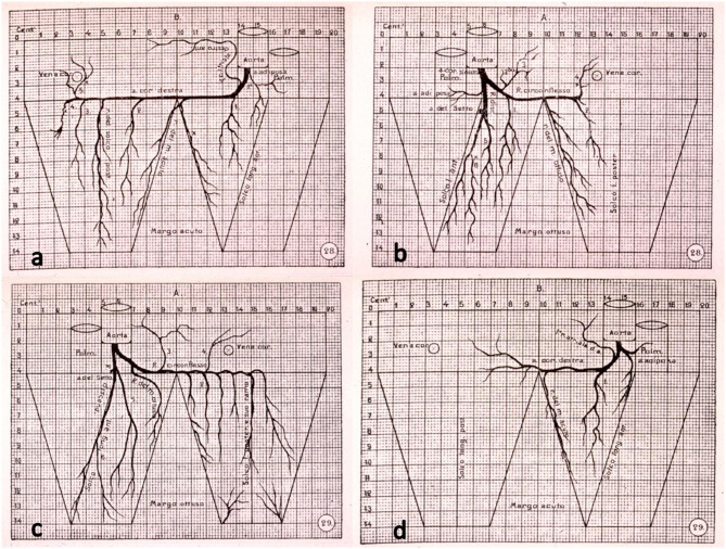
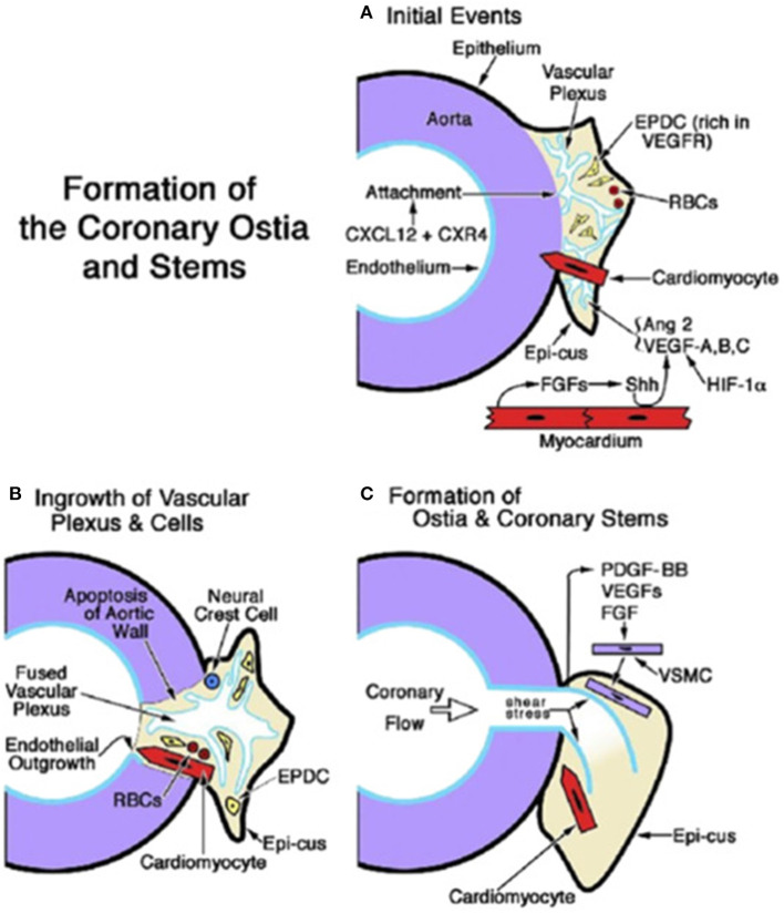
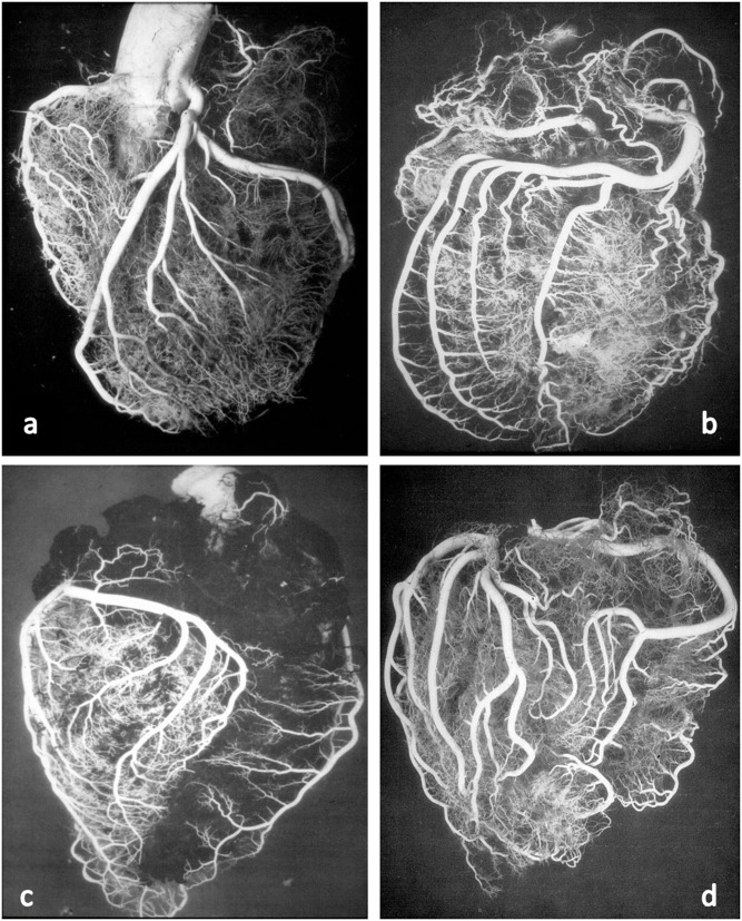
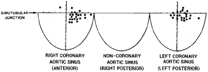
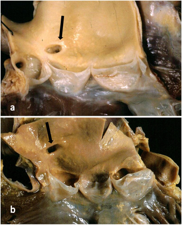
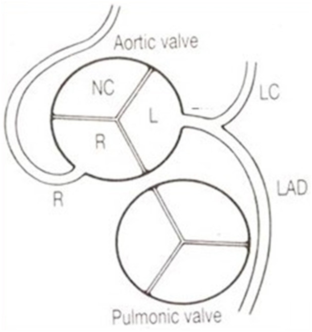
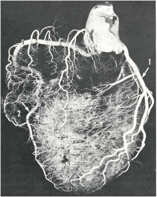
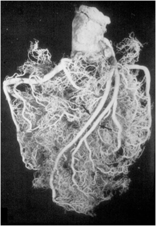
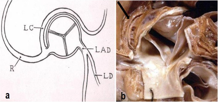
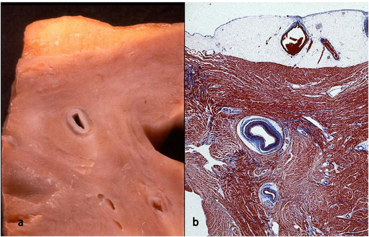
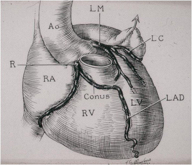
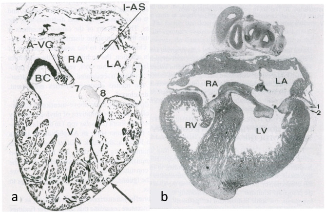
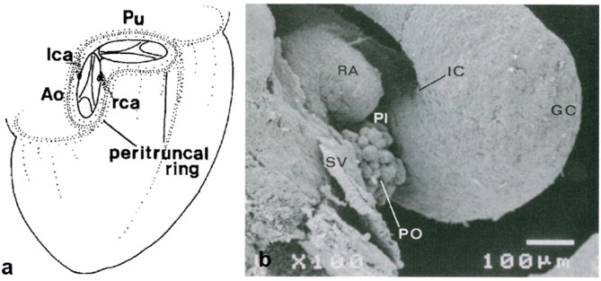
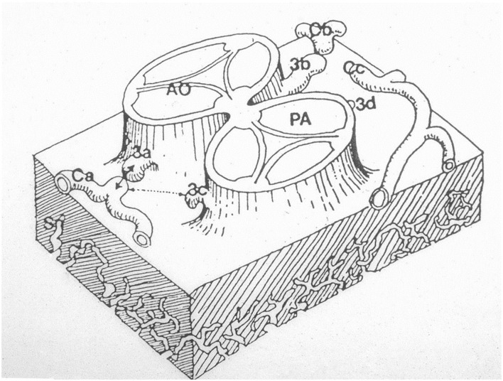
Anatomy of subepicardial coronary arteries became a topic of investigation at autopsy in Florence (Italy) by Banchi in the early twentieth century, with the discovery of dominant and balanced patterns. Thereafter, in the 60's of the same century Baroldi in Milan did post-mortem injection with spectacular three-dimensional casts. Later Sones at the Cleveland Clinic introduced selective coronary arteriography for visualization of coronary arteries. In the present chapter we show these patterns, as well as normal variants of origin and course with questionable risk of ischemia, like myocardial bridge as well as origin of the left circumflex coronary artery from the right sinus with retroaortic course. As far as embryology, the coronary arteries and veins are epicardial in origin and finally connect the former with the aorta, and the latter with the sinus venosus. At the time of spongy myocardium, intramural blood supply derives directly by the ventricular cavities, whereas later, at the time of myocardial compaction, vascularization originates from the subepicardial network. The connection of the subepicardial plexus with the aorta occurs with prongs of the peritruncal ring, which penetrate the facing aortic sinuses. Septation of truncus arteriosus is not responsible for the final position of the coronary orifices. Infact in transposition of the great arteries coronary ostia are regularly located within facing sinuses of the anterior aorta.
二十世纪初,意大利佛罗伦萨的班奇在尸检时将心外膜下冠状动脉的解剖结构作为研究课题,并发现了优势型和均衡型模式。此后,在同一世纪60年代,米兰的巴罗迪进行了尸检注射,制作出了壮观的三维铸型。后来,克利夫兰诊所的索内斯引入了选择性冠状动脉造影术来观察冠状动脉。在本章中,我们展示了这些模式,以及起源和走行的正常变异,这些变异存在可疑的缺血风险,如心肌桥以及左旋支冠状动脉从右窦起源并走行于主动脉后方。就胚胎学而言,冠状动脉和静脉起源于心外膜,最终前者与主动脉相连,后者与静脉窦相连。在海绵状心肌阶段,壁内血液供应直接来自心室腔,而后来,在心肌致密化阶段,血管化起源于心外膜下网络。心外膜下丛与主动脉的连接通过围绕动脉干环的分支实现,这些分支穿透相对的主动脉窦。动脉干的分隔与冠状动脉口的最终位置无关。事实上,在大动脉转位时,冠状动脉口通常位于前方主动脉相对的窦内。