Computational Neuroimaging Group, Trinity College Dublin, Dublin, Ireland
Computational Neuroimaging Group, Trinity College Dublin, Dublin, Ireland.
J Neurol Neurosurg Psychiatry. 2021 Nov;92(11):1197-1205. doi: 10.1136/jnnp-2021-326854. Epub 2021 Jun 24.
Cerebellar disease burden and cerebro-cerebellar connectivity alterations are poorly characterised in amyotrophic lateral sclerosis (ALS) despite the likely contribution of cerebellar pathology to the clinical heterogeneity of the condition.
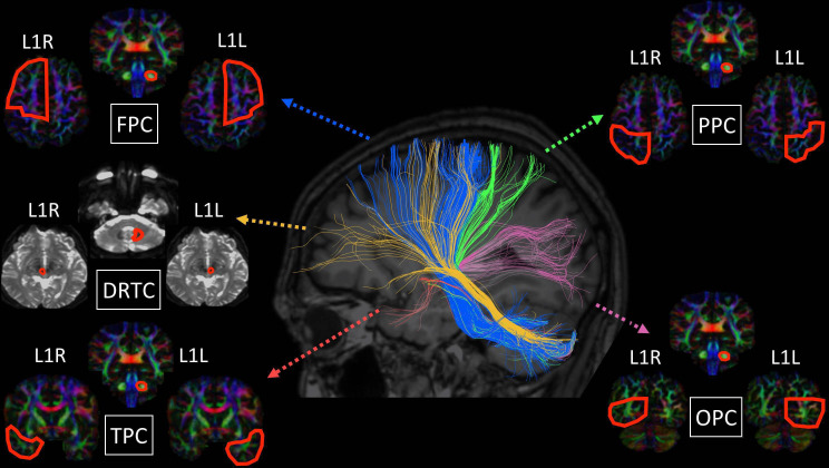
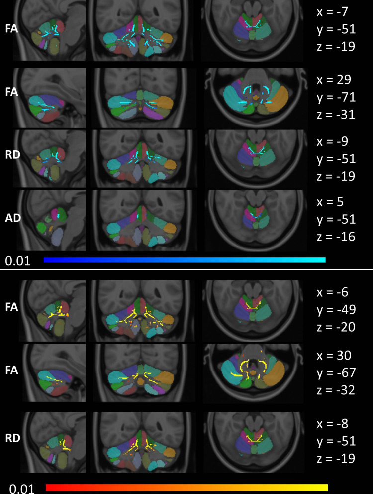
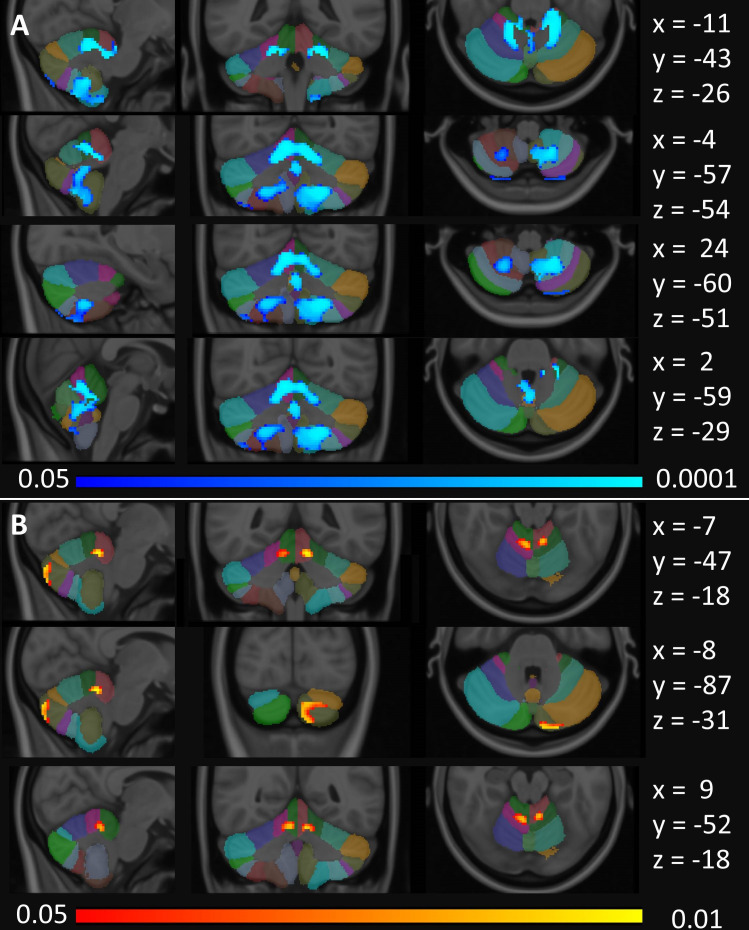
A prospective imaging study has been undertaken with 271 participants to systematically evaluate cerebellar grey and white matter alterations, cerebellar peduncle integrity and cerebro-cerebellar connectivity in ALS. Participants were stratified into four groups: (1) patients testing positive for GGGGCC repeat expansions in , (2) patients carrying an intermediate-length repeat expansion in , (3) patients without established ALS-associated mutations and (4) healthy controls. Additionally, the cerebellar profile of a single patient with ALS who had an allele length of 62 was evaluated. Cortical thickness, grey matter and white matter volumes were calculated in each cerebellar lobule complemented by morphometric analyses to characterise genotype-associated atrophy patterns. A Bayesian segmentation algorithm was used for superior cerebellar peduncle volumetry. White matter diffusivity parameters were appraised both within the cerebellum and in the cerebellar peduncles. Cerebro-cerebellar connectivity was assessed using deterministic tractography.
Cerebellar pathology was confined to lobules I-V of the anterior lobe in patients with sporadic ALS in contrast to the considerable posterior lobe and vermis disease burden identified in mutation carriers. Patients with intermediate expansions did not exhibit significant cerebellar pathology.
Focal rather than global cerebellar degeneration characterises ALS. Pathognomonic ALS symptoms which are typically attributed to other anatomical regions, such as dysarthria, dysphagia, pseudobulbar affect, eye movement abnormalities and cognitive deficits, may be modulated, exacerbated or partially driven by cerebellar changes in ALS.
尽管小脑病变可能导致肌萎缩侧索硬化症(ALS)临床表现的异质性,但小脑疾病负担和脑-小脑连接改变在 ALS 中仍描述不足。
对 271 名参与者进行了一项前瞻性影像学研究,以系统评估 ALS 患者小脑灰质和白质改变、小脑脚完整性和脑-小脑连接。参与者分为四组:(1)检测到 CAG 重复扩增的 ALS 患者,(2)携带中间长度重复扩增的 ALS 患者,(3)无明确 ALS 相关基因突变的 ALS 患者,(4)健康对照者。此外,还评估了一位携带 ALS 突变基因 等位基因长度为 62 的患者的小脑特征。对每个小脑叶进行皮质厚度、灰质和白质体积计算,并进行形态计量学分析以描述与基因型相关的萎缩模式。采用贝叶斯分割算法对小脑上脚进行体积测量。评估小脑内和小脑脚内的白质扩散参数。采用确定性追踪技术评估脑-小脑连接。
散发性 ALS 患者的小脑病变局限于前叶的Ⅰ-Ⅴ叶,而 突变携带者则存在明显的后叶和蚓部病变。中间长度 扩展的患者没有明显的小脑病变。
ALS 以局灶性而非全脑性小脑变性为特征。通常归因于其他解剖区域的 ALS 特有的症状,如构音障碍、吞咽困难、假性延髓性麻痹、眼球运动异常和认知缺陷,可能在 ALS 中被小脑改变所调节、加重或部分驱动。