From the Department of Neurology (H.K.v.d.B., H.-J.W., R.W., K.v.V., H.H.G.T., J.M.M., L.A.B., M.A.v.E., J.H.V., L.H.v.d.B.), Center of Excellence for Rehabilitation Medicine (L.A.B.), and Department of Radiology (J.H.), UMC Utrecht Brain Center, University Medical Center Utrecht; De Hoogstraat Rehabilitation (L.A.B.), Utrecht; and Department of Complex Trait Genetics (M.P.v.d.H.), Center for Neurogenomics and Cognitive Research, VU University Amsterdam, the Netherlands.
Neurology. 2020 Jun 16;94(24):e2592-e2604. doi: 10.1212/WNL.0000000000009498. Epub 2020 May 15.
To understand the progressive nature of amyotrophic lateral sclerosis (ALS) by investigating differential brain patterns of gray and white matter involvement in clinically or genetically defined subgroups of patients using cross-sectional, longitudinal, and multimodal MRI.
We assessed cortical thickness, subcortical volumes, and white matter connectivity from T1-weighted and diffusion-weighted MRI in 292 patients with ALS (follow-up: n = 150) and 156 controls (follow-up: n = 72). Linear mixed-effects models were used to assess changes in structural brain measurements over time in patients compared to controls.
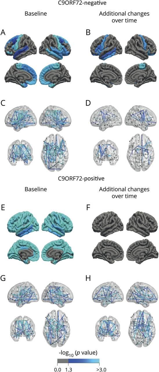
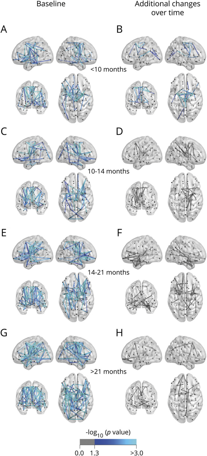
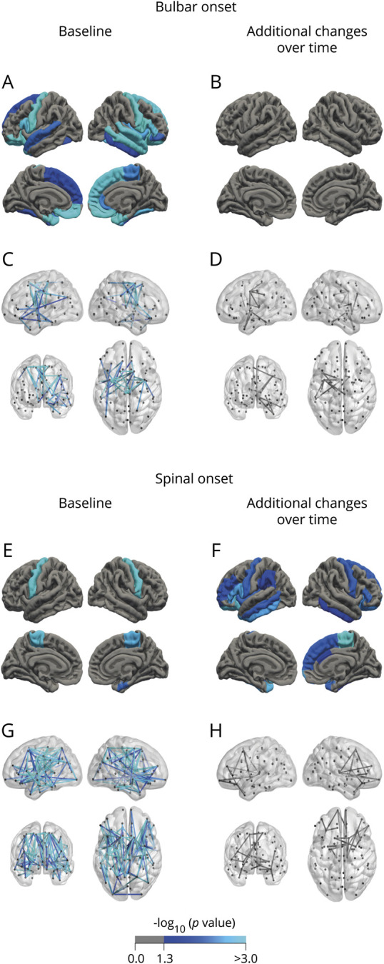
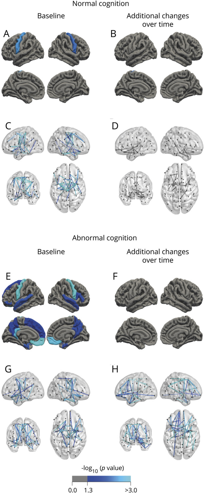
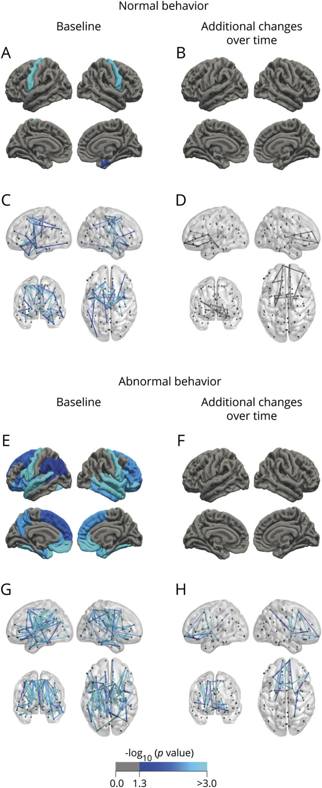
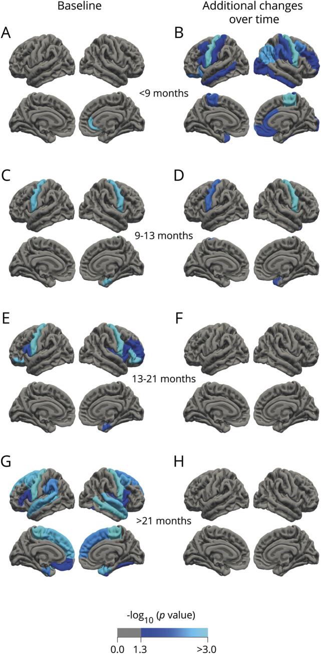
Patients with a mutation (n = 24) showed widespread gray and white matter involvement at baseline, and extensive loss of white matter integrity in the connectome over time. In -negative patients, we detected cortical thinning of motor and frontotemporal regions, and loss of white matter integrity of connections linked to the motor cortex. Patients with spinal onset displayed widespread white matter involvement at baseline and gray matter atrophy over time, whereas patients with bulbar onset started out with prominent gray matter involvement. Patients with unaffected cognition or behavior displayed predominantly motor system involvement, while widespread cerebral changes, including frontotemporal regions with progressive white matter involvement over time, were associated with impaired behavior or cognition. Progressive loss of gray and white matter integrity typically occurred in patients with shorter disease durations (<13 months), independent of progression rate.
Heterogeneity of phenotype and genotype relates to distinct patterns of cerebral degeneration. We demonstrate that imaging studies have the potential to monitor disease progression and early intervention may be required to limit cerebral degeneration.
通过对临床或基因定义的患者亚组进行横断面、纵向和多模态 MRI 研究,探讨灰质和白质受累的差异脑模式,了解肌萎缩侧索硬化症(ALS)的进展性。
我们评估了 292 名 ALS 患者(随访:n = 150)和 156 名对照者(随访:n = 72)的 T1 加权和弥散加权 MRI 的皮质厚度、皮质下体积和白质连接。线性混合效应模型用于评估与对照组相比,患者的结构脑测量值随时间的变化。
携带 突变的患者(n = 24)在基线时表现出广泛的灰质和白质受累,并随时间推移连接组中白质完整性的广泛丧失。在 -阴性患者中,我们检测到运动和额颞叶区域的皮质变薄,以及与运动皮层相连的白质完整性的丧失。脊髓起病的患者在基线时表现出广泛的白质受累和随时间推移的灰质萎缩,而延髓起病的患者则以明显的灰质受累为首发症状。认知或行为不受影响的患者主要表现为运动系统受累,而广泛的大脑变化,包括额颞叶区域随时间推移逐渐出现的白质受累,与行为或认知障碍有关。灰质和白质完整性的进行性丧失通常发生在疾病持续时间较短(<13 个月)的患者中,与进展速度无关。
表型和基因型的异质性与不同的大脑退化模式有关。我们证明,影像学研究具有监测疾病进展的潜力,可能需要早期干预来限制大脑退化。