• 文献检索
  • 文档翻译
  • 深度研究
  • 学术资讯
  • Suppr Zotero 插件Zotero 插件
  • 邀请有礼
  • 套餐&价格
  • 历史记录
应用&插件
Suppr Zotero 插件Zotero 插件浏览器插件Mac 客户端Windows 客户端微信小程序
定价
高级版会员购买积分包购买API积分包
服务
文献检索文档翻译深度研究API 文档MCP 服务
关于我们
关于 Suppr公司介绍联系我们用户协议隐私条款
关注我们

Suppr 超能文献

核心技术专利:CN118964589B侵权必究
粤ICP备2023148730 号-1Suppr @ 2026

文献检索

告别复杂PubMed语法,用中文像聊天一样搜索,搜遍4000万医学文献。AI智能推荐,让科研检索更轻松。

立即免费搜索

文件翻译

保留排版,准确专业,支持PDF/Word/PPT等文件格式,支持 12+语言互译。

免费翻译文档

深度研究

AI帮你快速写综述,25分钟生成高质量综述,智能提取关键信息,辅助科研写作。

立即免费体验

[磁共振成像在腮腺肿瘤组织病理学中的表现]

[Performance of the magnetic resonance imaging in parotid gland tumor histopathology].

作者信息

Masmoudi Mohamed, Hasnaoui Mehdi, Guizani Rihab, Lahmar Rihab, Jerbi Saida, Mighri Khalifa

机构信息

Département d´Oto-rhino-laryngologie, Hôpital Tahar Sfar Mahdia, Mahdia, Tunisia.

Département d´Imagerie Médicale, Hôpital Tahar Sfar Mahdia, Mahdia, Tunisia.

出版信息

Pan Afr Med J. 2021 May 4;39:10. doi: 10.11604/pamj.2021.39.10.27813. eCollection 2021.

DOI:10.11604/pamj.2021.39.10.27813
PMID:34178238
原文链接:https://pmc.ncbi.nlm.nih.gov/articles/PMC8197060/
Abstract

INTRODUCTION

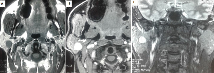
salivary gland tumors mainly occur in the parotid gland. These tumors are rare but are characterized by histological heterogeneity, thus posing diagnostic challenges. Magnetic resonance imaging (MRI) is currently the most reliable imaging test for the evaluation of these tumors. The purpose of this study was to highlight the diagnostic value of MRI and its role in parotid gland tumor histopathology.

METHODS

we conducted a retrospective descriptive and analytical study of 50 patients with parotid gland tumor, operated and treated in the ear, nose and throat (ENT) Department and in the Department of cervicofacial surgery at the Tahar Sfar University Hospital of Mahdia between 2001 and 2019. All patients underwent preoperative MRI of the parotid gland.

RESULTS

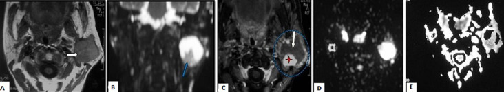
out of 50 patients included in the study, 36 (72%) had benign tumor and 14 (28%) malignant tumor. The sensitivity of MRI for the diagnosis of malignant tumor was 92.8% with a specificity of 97.2%, a negative predictive value of 93% and a positive predictive value of 97%. With respect to benign tumor characterization, MRI suggested the diagnosis of Warthin tumor in all cases (13 cases) and of pleomorphic adenoma in 22 out of 23 cases. There were two diagnostic errors: MRI suggested the diagnosis of pleomorphic adenoma instead of adenoid cystic carcinoma in one case and of malignant tumor instead of pleomorphic adenoma due to diffusion restriction.

CONCLUSION

MRI is highly efficient in the assessment of parotid tumor histology and, especially, after the advent of new functional sequences. However, only histological examination allows to confirm with certainty the diagnosis.

摘要

引言

唾液腺肿瘤主要发生于腮腺。这些肿瘤较为罕见,但具有组织学异质性,因此带来了诊断挑战。磁共振成像(MRI)是目前评估这些肿瘤最可靠的影像学检查。本研究的目的是突出MRI的诊断价值及其在腮腺肿瘤组织病理学中的作用。

方法

我们对2001年至2019年期间在马赫迪耶塔哈尔·斯法尔大学医院耳鼻喉科(ENT)和颈面部外科接受手术治疗的50例腮腺肿瘤患者进行了回顾性描述性和分析性研究。所有患者均接受了腮腺术前MRI检查。

结果

在纳入研究的50例患者中,36例(72%)为良性肿瘤,14例(28%)为恶性肿瘤。MRI诊断恶性肿瘤的敏感性为92.8%,特异性为97.2%,阴性预测值为93%,阳性预测值为97%。关于良性肿瘤的特征,MRI在所有病例(13例)中提示了沃辛瘤的诊断,在23例中的22例中提示了多形性腺瘤的诊断。有两例诊断错误:一例中MRI提示为多形性腺瘤而非腺样囊性癌,另一例因扩散受限提示为恶性肿瘤而非多形性腺瘤。

结论

MRI在评估腮腺肿瘤组织学方面非常高效,尤其是在新的功能序列出现之后。然而,只有组织学检查才能确定确诊。

https://cdn.ncbi.nlm.nih.gov/pmc/blobs/1a15/8197060/dc89c07ed853/PAMJ-39-10-g002.jpg
https://cdn.ncbi.nlm.nih.gov/pmc/blobs/1a15/8197060/556da4f09295/PAMJ-39-10-g001.jpg
https://cdn.ncbi.nlm.nih.gov/pmc/blobs/1a15/8197060/dc89c07ed853/PAMJ-39-10-g002.jpg
https://cdn.ncbi.nlm.nih.gov/pmc/blobs/1a15/8197060/556da4f09295/PAMJ-39-10-g001.jpg
https://cdn.ncbi.nlm.nih.gov/pmc/blobs/1a15/8197060/dc89c07ed853/PAMJ-39-10-g002.jpg

相似文献

1
[Performance of the magnetic resonance imaging in parotid gland tumor histopathology].[磁共振成像在腮腺肿瘤组织病理学中的表现]
Pan Afr Med J. 2021 May 4;39:10. doi: 10.11604/pamj.2021.39.10.27813. eCollection 2021.
2
Value of T2-weighted-based radiomics model in distinguishing Warthin tumor from pleomorphic adenoma of the parotid.基于 T2 加权的放射组学模型在鉴别腮腺沃辛瘤与多形性腺瘤中的价值。
Eur Radiol. 2023 Jun;33(6):4453-4463. doi: 10.1007/s00330-022-09295-0. Epub 2022 Dec 11.
3
Diagnostic performance of qualitative and radiomics approach to parotid gland tumors: which is the added benefit of texture analysis?定性和影像组学方法诊断腮腺肿瘤的性能:纹理分析的附加益处是什么?
Br J Radiol. 2021 Dec;94(1128):20210340. doi: 10.1259/bjr.20210340. Epub 2021 Sep 30.
4
An ultrasound-based ensemble machine learning model for the preoperative classification of pleomorphic adenoma and Warthin tumor in the parotid gland.基于超声的集成机器学习模型用于术前腮腺多形性腺瘤和沃辛瘤的分类。
Eur Radiol. 2024 Oct;34(10):6862-6876. doi: 10.1007/s00330-024-10719-2. Epub 2024 Apr 3.
5
Parotid gland tumors: comparison of conventional and diffusion-weighted MRI findings with histopathological results.腮腺肿瘤:常规 MRI 与弥散加权 MRI 表现与组织病理学结果的对比研究。
Dentomaxillofac Radiol. 2021 May 1;50(4):20200391. doi: 10.1259/dmfr.20200391. Epub 2020 Dec 11.
6
Clinical and demographic data improve diagnostic accuracy of dynamic contrast-enhanced and diffusion-weighted MRI in differential diagnostics of parotid gland tumors.临床和人口统计学数据可提高动态对比增强和弥散加权 MRI 在腮腺肿瘤鉴别诊断中的诊断准确性。
Oral Oncol. 2020 Dec;111:104932. doi: 10.1016/j.oraloncology.2020.104932. Epub 2020 Jul 30.
7
Do magnetic resonance imaging and ultrasound add anything to the preoperative workup of parotid gland tumors?磁共振成像和超声检查对腮腺肿瘤的术前检查有何补充作用?
J Oral Maxillofac Surg. 2007 May;65(5):945-52. doi: 10.1016/j.joms.2006.04.046.
8
Imaging quality of PROPELLER diffusion-weighted MR imaging and its diagnostic performance in distinguishing pleomorphic adenomas from Warthin tumors of the parotid gland.螺旋桨扩散加权磁共振成像的成像质量及其对鉴别腮腺多形性腺瘤和沃辛瘤的诊断性能。
NMR Biomed. 2020 May;33(5):e4282. doi: 10.1002/nbm.4282. Epub 2020 Mar 2.
9
[Correlation between magnetic resonance imaging and extemporaneous and definitive histological examination of parotid gland tumors: a case series].[腮腺肿瘤的磁共振成像与即时和确定性组织学检查之间的相关性:病例系列]
Pan Afr Med J. 2020 Sep 22;37:80. doi: 10.11604/pamj.2020.37.80.21192. eCollection 2020.
10
T2 mapping and readout segmentation of long variable echo-train diffusion-weighted imaging for the differentiation of parotid gland tumors.用于腮腺肿瘤鉴别诊断的长可变回波链扩散加权成像的T2映射和读出分割
Eur J Radiol. 2022 Jun;151:110265. doi: 10.1016/j.ejrad.2022.110265. Epub 2022 Mar 25.

引用本文的文献

1
Diagnosis of parotid gland tumors using a ternary classification model based on ultrasound radiomics.基于超声影像组学的腮腺肿瘤三元分类模型诊断
Front Oncol. 2025 Mar 21;15:1485393. doi: 10.3389/fonc.2025.1485393. eCollection 2025.
2
Diagnostic performance of MRI-based radiomics models using machine learning approaches for the triple classification of parotid tumors.使用机器学习方法的基于MRI的放射组学模型对腮腺肿瘤进行三重分类的诊断性能。
Heliyon. 2024 Aug 22;10(17):e36601. doi: 10.1016/j.heliyon.2024.e36601. eCollection 2024 Sep 15.
3
Utility of fine-needle aspiration biopsy (FNAB) in parotid pleomorphic adenoma diagnosis and management.

本文引用的文献

1
[Correlation between magnetic resonance imaging and extemporaneous and definitive histological examination of parotid gland tumors: a case series].[腮腺肿瘤的磁共振成像与即时和确定性组织学检查之间的相关性:病例系列]
Pan Afr Med J. 2020 Sep 22;37:80. doi: 10.11604/pamj.2020.37.80.21192. eCollection 2020.
2
Sensitivity of Fine-Needle Aspiration and Imaging Modalities in the Diagnosis of Low-Grade Mucoepidermoid Carcinoma of the Parotid Gland.细针穿刺活检及影像学检查在腮腺低度黏液表皮样癌诊断中的敏感性
Ann Otol Rhinol Laryngol. 2019 Aug;128(8):755-759. doi: 10.1177/0003489419842582. Epub 2019 Apr 16.
3
细针穿刺活检(FNAB)在腮腺多形性腺瘤诊断和治疗中的应用。
Biomed Pap Med Fac Univ Palacky Olomouc Czech Repub. 2024 Jun;168(2):156-161. doi: 10.5507/bp.2023.027. Epub 2023 Jul 10.
4
A Deep Learning Model for Classification of Parotid Neoplasms Based on Multimodal Magnetic Resonance Image Sequences.基于多模态磁共振图像序列的腮腺肿瘤分类深度学习模型。
Laryngoscope. 2023 Feb;133(2):327-335. doi: 10.1002/lary.30154. Epub 2022 May 16.
Contemporary Management of Benign and Malignant Parotid Tumors.
腮腺良恶性肿瘤的当代管理
Front Surg. 2018 May 11;5:39. doi: 10.3389/fsurg.2018.00039. eCollection 2018.
4
Cross-sectional Imaging of Parotid Gland Nodules: A Brief Practical Guide.腮腺结节的横断面成像:简要实用指南
J Clin Imaging Sci. 2018 Apr 16;8:14. doi: 10.4103/jcis.JCIS_8_18. eCollection 2018.
5
Analysis of efficacy and safety of core-needle biopsy versus fine-needle aspiration cytology in patients with cervical lymphadenopathy and salivary gland tumour.核心针吸活检与细针穿刺细胞学检查在颈部淋巴结病和涎腺肿瘤患者中的疗效及安全性分析
Int J Oral Maxillofac Surg. 2018 Oct;47(10):1229-1235. doi: 10.1016/j.ijom.2018.04.003. Epub 2018 Apr 27.
6
WHO is in and WHO is out of the mouth, salivary glands, and jaws sections of the 4th edition of the WHO classification of head and neck tumours.在《世界卫生组织头颈部肿瘤分类》第4版中,口腔、唾液腺及颌部章节所涵盖和未涵盖的内容。
Br J Oral Maxillofac Surg. 2018 Feb;56(2):90-95. doi: 10.1016/j.bjoms.2017.12.009. Epub 2018 Jan 17.
7
[Cyto-histological correlation in the management of parotid gland tumors: A retrospective study of 160 cases].腮腺肿瘤治疗中的细胞组织学相关性:160例回顾性研究
Bull Cancer. 2017 Oct;104(10):850-857. doi: 10.1016/j.bulcan.2017.09.001. Epub 2017 Oct 12.
8
Diffusion- and Perfusion-Weighted Magnetic Resonance Imaging-An Alternative to Fine Needle Biopsy or Only an Adjunct Test in Preoperative Differential Diagnostics of Malignant and Benign Parotid Tumors?扩散加权磁共振成像和灌注加权磁共振成像——细针穿刺活检的替代方法,还是仅作为腮腺良恶性肿瘤术前鉴别诊断的辅助检查?
J Oral Maxillofac Surg. 2017 Oct;75(10):2248-2253. doi: 10.1016/j.joms.2017.03.018. Epub 2017 Mar 22.
9
The accuracy of fine-needle aspiration cytology for diagnosis of parotid gland masses: a clinicopathological study of 114 patients.细针穿刺细胞学检查诊断腮腺肿块的准确性:114例患者的临床病理研究
J Appl Oral Sci. 2016 Nov-Dec;24(6):561-567. doi: 10.1590/1678-775720160214.
10
[Predictors of malignancy in the management of parotid tumors: about 76 cases].腮腺肿瘤治疗中恶性肿瘤的预测因素:约76例报告
Pan Afr Med J. 2016 Mar 16;23:112. doi: 10.11604/pamj.2016.23.112.8404. eCollection 2016.