Tang Shiyuan, Fan Chengming, Iroegbu Chukwuemeka Daniel, Zhou Wenwu, Zhang Zhigong, Wu Ming, Chen Wangping, Wu Xiaoming, Peng Jun, Li Zhihong, Yang Jinfu
Department of the Cardiovascular Surgery, The Second Xiangya Hospital, Central South University, Changsha, China.
Department of the Cardiovascular Surgery of the Hunan Provincial People's Hospital, Changsha, China.
Front Cell Dev Biol. 2021 Jun 9;9:670913. doi: 10.3389/fcell.2021.670913. eCollection 2021.
The actin-sequestering proteins, thymosin beta-4 (Tβ4) and hypoxia-inducible factor (HIF)-1α, are known to be associated with angiogenesis after myocardial infarction (MI). Herein, we aimed to identify the mechanism of HIF-1α induction by Tβ4 and investigate the effects of bone marrow mesenchymal stromal cells (BMMSCs) transfected with the Tβ4 gene () in a rat model of MI.
Rat BMMSCs were isolated, cultured, and transfected with the gene by using the lentivirus-mediated method. Rats with surgically induced MI were randomly divided into three groups ( = 9/group); after 1 week, the rats were injected at the heart infarcted border zone with TMSB4-overexpressed BMMSCs (BMMSC-TMSB4 ), wild-type BMMSCs that expressed normal levels of TMSB4 (BMMSC-TMSB4 ), or medium (MI). The fourth group of animals ( = 9) underwent all surgical procedures necessary for MI induction except for the ligation step (Sham). Four weeks after the injection, heart function was measured using transthoracic echocardiography. Infarct size was calculated by TTC staining, and collagen volume was measured by Masson staining. Angiogenesis in the infarcted heart area was evaluated by CD31 immunofluorescence histochemistry. experiments were carried out to observe the effect of exogenous Tβ4 on HIF-1α and explore the various possible mechanism(s).
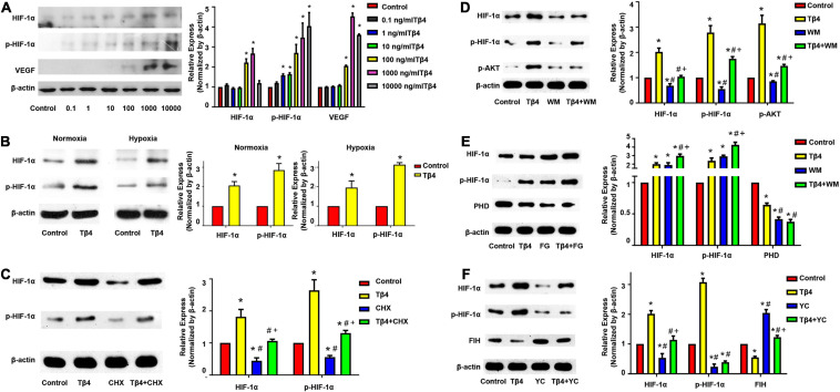
experiments showed that vascular density 4 weeks after treatment was about twofold higher in BMMSC-TMSB4 -treated animals than in BMMSC-TMSB4 -treated animals ( < 0.05). The cardiac function and infarct size significantly improved in both cell-treatment groups compared to controls. Notably, the cardiac function and infarct size were most prominent in BMMSC-TMSB4 -treated animals (both < 0.05). HIF-1α and phosphorylated HIF-1α (p-HIF-1α) were significantly enhanced by exogenous Tβ4, which was nonetheless blocked by the factor-inhibiting HIF (FIH) promoter (YC-1). The expression of prolyl hydroxylase domain proteins (PHD) was decreased upon treatment with Tβ4 and further decreased with the combined treatment of Tβ4 and FG-4497 (a specific PHD inhibitor).
TMSB4-transfected BMMSCs might significantly improve recovery from myocardial ischemia and promote the generation of HIF-1α and p-HIF-1α the AKT pathway, and inhibit the degradation of HIF-1α the PHD and FIH pathways.
已知肌动蛋白隔离蛋白胸腺素β-4(Tβ4)和缺氧诱导因子(HIF)-1α与心肌梗死(MI)后的血管生成有关。在此,我们旨在确定Tβ4诱导HIF-1α的机制,并研究用Tβ4基因转染的骨髓间充质基质细胞(BMMSCs)在大鼠MI模型中的作用。
分离、培养大鼠BMMSCs,并采用慢病毒介导的方法用该基因转染。将手术诱导MI的大鼠随机分为三组(每组n = 9);1周后,在心脏梗死边缘区注射过表达TMSB4的BMMSCs(BMMSC-TMSB4 )、表达正常水平TMSB4的野生型BMMSCs(BMMSC-TMSB4 )或培养基(MI组)。第四组动物(n = 9)除结扎步骤外,接受诱导MI所需的所有手术操作(假手术组)。注射后4周,采用经胸超声心动图测量心脏功能。通过TTC染色计算梗死面积,通过Masson染色测量胶原体积。通过CD31免疫荧光组织化学评估梗死心脏区域的血管生成。进行实验以观察外源性Tβ4对HIF-1α的影响,并探索各种可能的机制。
实验表明,治疗4周后,BMMSC-TMSB4 治疗组动物的血管密度比BMMSC-TMSB4 治疗组动物高约两倍(P < 0.05)。与对照组相比,两个细胞治疗组的心脏功能和梗死面积均显著改善。值得注意的是,BMMSC-TMSB4 治疗组动物的心脏功能和梗死面积改善最为显著(均P < 0.05)。外源性Tβ4显著增强了HIF-1α和磷酸化HIF-1α(p-HIF-1α),但被缺氧诱导因子抑制因子(FIH)启动子(YC-1)阻断。用Tβ4处理后脯氨酰羟化酶结构域蛋白(PHD)的表达降低,Tβ4与FG-4497(一种特异性PHD抑制剂)联合处理后进一步降低。
转染TMSB4的BMMSCs可能显著改善心肌缺血后的恢复,并通过AKT途径促进HIF-1α和p-HIF-1α的生成,通过PHD和FIH途径抑制HIF-1α的降解。