Department of Neurobiology, Duke University Medical Center.
Department of Neurobiology, Duke University Medical Center;
J Vis Exp. 2021 Jun 9(172). doi: 10.3791/62568.
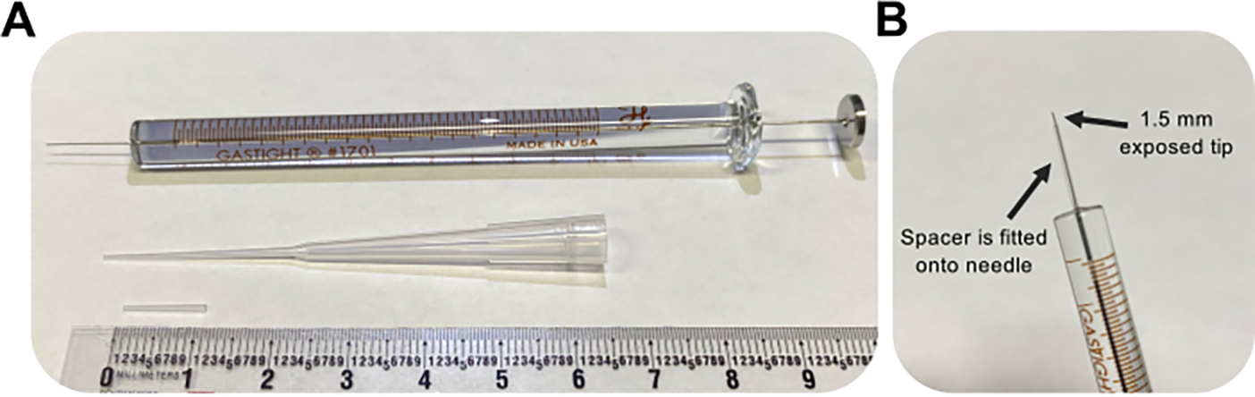
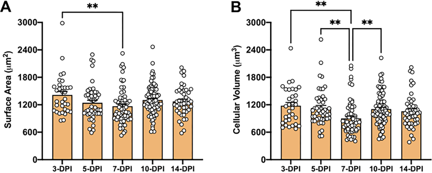
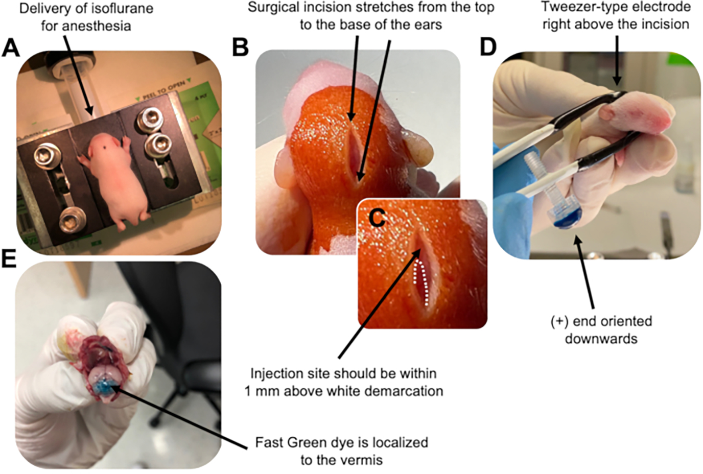
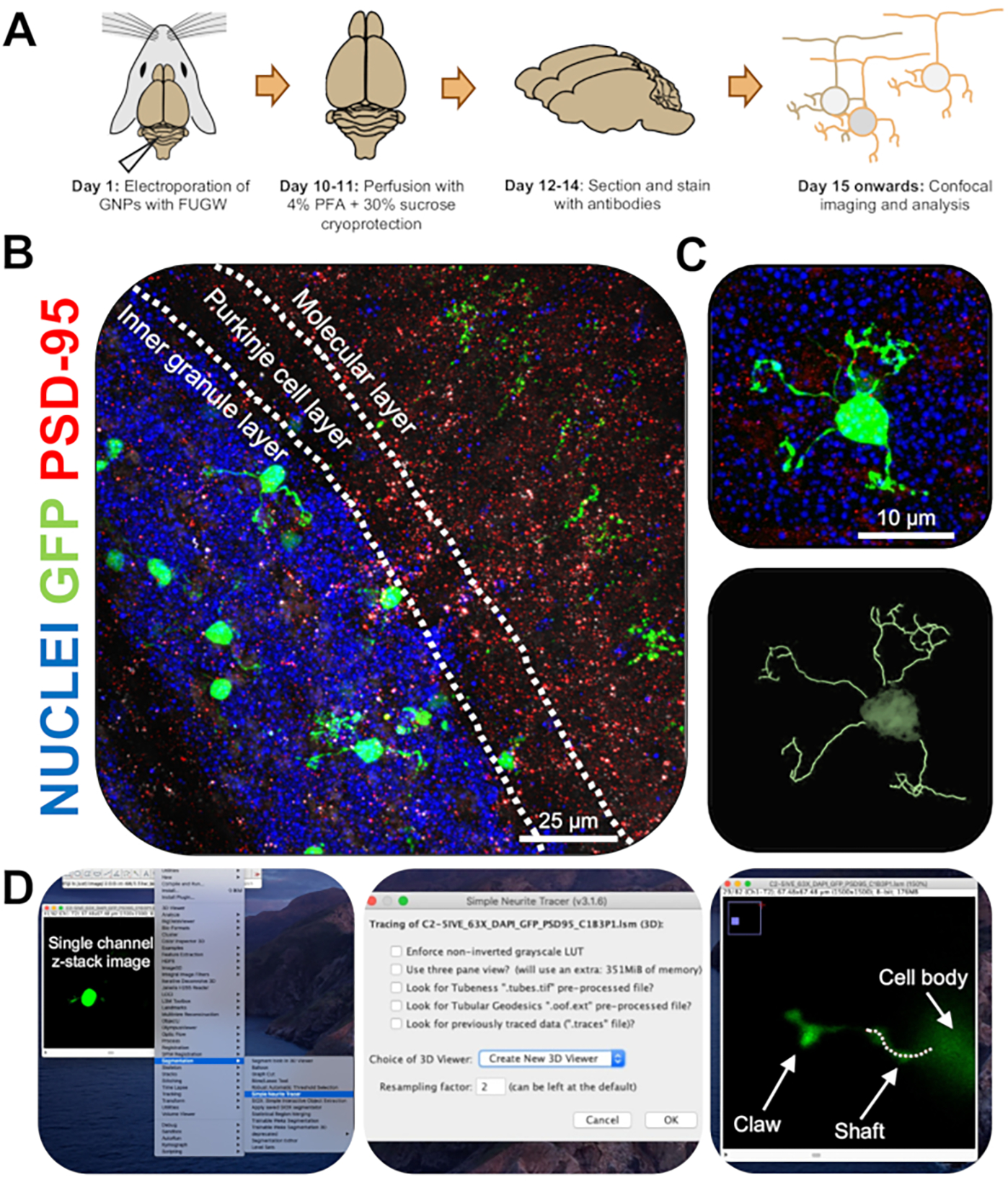
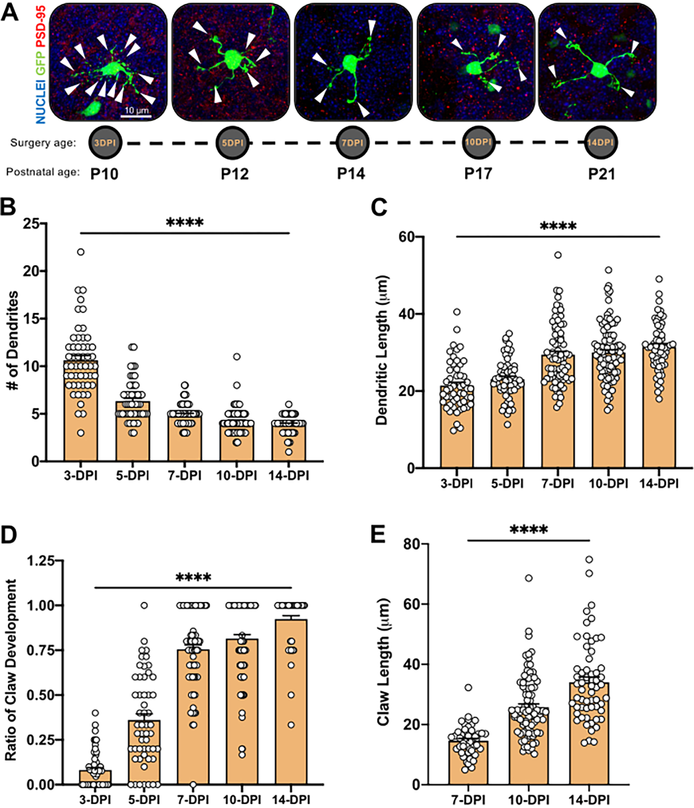
Neurons undergo dynamic changes in their structure and function during brain development to form appropriate connections with other cells. The rodent cerebellum is an ideal system to track the development and morphogenesis of a single cell type, the cerebellar granule neuron (CGN), across time. Here, in vivo electroporation of granule neuron progenitors in the developing mouse cerebellum was employed to sparsely label cells for subsequent morphological analyses. The efficacy of this technique is demonstrated in its ability to showcase key developmental stages of CGN maturation, with a specific focus on the formation of dendritic claws, which are specialized structures where these cells receive the majority of their synaptic inputs. In addition to providing snapshots of CGN synaptic structures throughout cerebellar development, this technique can be adapted to genetically manipulate granule neurons in a cell-autonomous manner to study the role of any gene of interest and its effect on CGN morphology, claw development, and synaptogenesis.
在大脑发育过程中,神经元的结构和功能会发生动态变化,以与其他细胞形成适当的连接。啮齿动物小脑是一个理想的系统,可以追踪单个细胞类型(小脑颗粒神经元,CGN)随时间的发育和形态发生。在这里,通过在发育中的小鼠小脑内对颗粒神经元前体细胞进行活体电穿孔,稀疏标记细胞以进行后续的形态分析。该技术的有效性体现在其能够展示 CGN 成熟的关键发育阶段,特别关注树突爪的形成,这是这些细胞接收其大部分突触输入的专门结构。除了提供整个小脑发育过程中 CGN 突触结构的快照外,该技术还可以适应以细胞自主的方式对颗粒神经元进行遗传操作,以研究任何感兴趣基因的作用及其对 CGN 形态、爪发育和突触发生的影响。