Dean Benjamin J F, Little Christopher, Riley Nicholas D, Sellon Edward, Sheehan Warren, Burford Jenna, Hormbrey Phil, Costa Matthew L
Nuffield Department of Orthopaedics, Rheumatology and Musculoskeletal Sciences, University of Oxford, Oxford, UK.
Department of Orthopaedics, Oxford University Hospitals NHS Foundation Trust Nuffield Orthopaedic Centre, Oxford, UK.
Bone Jt Open. 2021 Jun;2(6):447-453. doi: 10.1302/2633-1462.26.BJO-2021-0054.R1.
To determine the role of early MRI in the management of suspected scaphoid fractures.
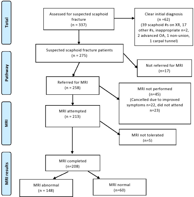
A total of 337 consecutive patients presenting to an emergency department (ED) following wrist trauma over a 12-month period were prospectively included in this service evaluation project. MRI was not required in 62 patients with clear diagnoses, and 17 patients were not managed as per pathway, leaving a total of 258 patients with normal scaphoid series radiographs who were then referred directly from ED for an acute wrist MRI scan. Patient demographics, clinical details, outcomes, and complications were recorded at a minimum of a year following injury.
The median time from injury to ED presentation was one day and the median number of positive clinical signs was two out of three (snuffbox tenderness, tubercle tenderness, pain on telescoping). Of 258 patients referred for acute MRI, 208 scans were performed as 50 patients either did not tolerate (five patients) or did not attend their scan (45 patients). MRI scans demonstrated scaphoid fracture (13%), fracture of another bone (22%), scaphoid contusion (6%), other contusion/ligamentous injury (20%), or solely degenerative pathology (10%). Only 29% of scans showed no abnormality. Almost 50% of those undergoing MRI (100 patients) were discharged by ED with advice, with only one re-presentation. Of the 27 undisplaced occult scaphoid fractures, despite prompt cast immobilization, two experienced delayed union which was successfully treated with surgery.
The use of MRI direct from ED enables prompt diagnosis and the early discharge of a large proportion of patients with normal radiographs following wrist trauma. Cite this article: 2021;2(6):447-453.
确定早期磁共振成像(MRI)在疑似舟骨骨折治疗中的作用。
在一项服务评估项目中,前瞻性纳入了在12个月期间因腕部创伤到急诊科就诊的337例连续患者。62例诊断明确的患者无需进行MRI检查,17例患者未按流程处理,剩余258例舟骨系列X线片正常的患者随后直接从急诊科转诊进行急性腕部MRI扫描。在受伤至少一年后记录患者的人口统计学资料、临床细节、治疗结果和并发症。
从受伤到到急诊科就诊的中位时间为1天,阳性临床体征的中位数为三项中的两项(鼻烟窝压痛、结节压痛、纵向挤压痛)。在转诊进行急性MRI检查的258例患者中,208例进行了扫描,50例患者因不耐受(5例)或未参加扫描(45例)而未进行检查。MRI扫描显示舟骨骨折(13%)、其他骨骼骨折(22%)、舟骨挫伤(6%)、其他挫伤/韧带损伤(20%)或仅有退行性病变(10%)。只有29%的扫描未显示异常。接受MRI检查的患者中近50%(100例)在急诊科接受建议后出院,仅1例再次就诊。在27例无移位的隐匿性舟骨骨折中,尽管及时进行了石膏固定,仍有2例出现延迟愈合,经手术成功治疗。
直接从急诊科使用MRI能够对腕部创伤后X线片正常的大部分患者进行快速诊断和早期出院。引用本文:2021;2(6):447 - 453。