Maxillofacial Surgery - Hospital of Treviso, Treviso, Italy.
Anatomical Pathology - Hospital of Treviso, Treviso, Italy.
J Oral Rehabil. 2021 Sep;48(9):1025-1034. doi: 10.1111/joor.13218. Epub 2021 Jul 9.
The aim of this study is to show the anatomical and histological features of the displaced temporomandibular joint (TMJ) disc in joints with degenerative disease.
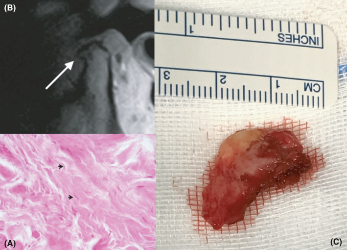
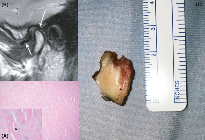
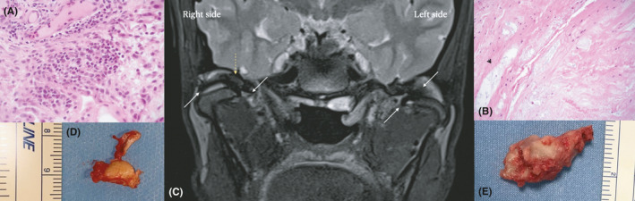
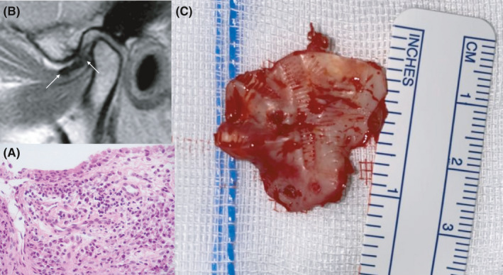
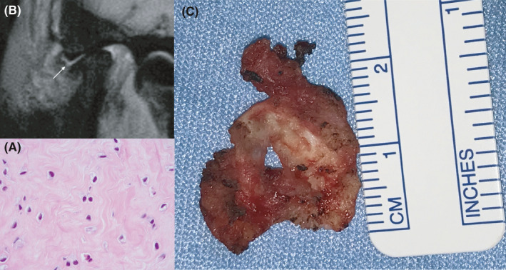
This study was performed on a total of 30 TMJ discs extracted from 22 patients, who underwent surgical discectomy after failure of conservative non-surgical treatment regimens to control pain and/or limited range of motion. All joints had imaging signs of an anteriorized disc position and degenerative joint disease. Samples of the extracted discs were stored in formalin, cut into 3 micron-thick sections imbedded in paraffin and processed with hematoxylin-eosin.
All the samples present irreversible morphologic and histological alterations. The macroscopical evaluation showed that 14 discs were worn and fragmented in several parts, and one disc was perforated. Morphological alterations with deformation and degenerative signs were shown in all discs, which were all severely worn and compromised. Histologically, various alterations were found, such as pre-fibrous sclerosis with myxoid degeneration and collagen deposits (N = 25), an increase in fibro-hyaline and fibrous tissues, with loss of elasticity (N = 25), scattered calcifications (N = 15), and synovial inflammation with microvascular proliferation and increased cellularity, presence of lymphocytes, histiocytes and plasma cells (N = 18). After the intervention, all patients reported decreased pain levels and showed improved function at 6 months.
These observations suggest that degenerative joint disease is accompanied by a anteriorized discs featuring abnormal macroscopical and histological changes. From a clinical viewpoint, this may suggest that, when treatment escalation leads to consider TMJ surgery, total discectomy is the most reasonable approach.
本研究旨在展示退行性疾病关节中移位的颞下颌关节(TMJ)盘的解剖和组织学特征。
本研究共纳入 22 名患者的 30 个 TMJ 盘,这些患者在保守的非手术治疗方案失败后,因疼痛和/或运动范围受限而行手术关节盘切除术。所有关节均有影像学表现为盘前移和退行性关节病。提取的关节盘样本用福尔马林保存,切成 3 微米厚的石蜡包埋切片,并用苏木精-伊红处理。
所有样本均呈现不可逆的形态和组织学改变。宏观评估显示,14 个关节盘磨损和碎裂成几个部分,1 个关节盘穿孔。所有关节盘均显示形态改变和退行性改变的迹象,均严重磨损和受损。组织学上发现各种改变,如纤维前硬化伴黏液样变性和胶原沉积(N=25),纤维-透明质酸和纤维组织增加,弹性丧失(N=25),散在钙化(N=15),以及滑膜炎症伴微血管增生和细胞增多,淋巴细胞、组织细胞和浆细胞存在(N=18)。干预后,所有患者均报告疼痛水平降低,6 个月时功能改善。
这些观察结果表明退行性关节病伴有盘前移,其特征为异常的宏观和组织学改变。从临床角度来看,这可能表明,当治疗升级导致考虑 TMJ 手术时,全关节盘切除术是最合理的方法。