Fukunaga Kanako, Tanji Masafumi, Hanzawa Nana, Kuroda Hiroki, Inui Masafumi
Systems Biology Program, Graduate School of Media and Governance, Keio University, Kanagawa, 252-0882, Japan.
Institute for Advanced Biosciences, Keio University, Kanagawa, 252-0882, Japan.
Biochem Biophys Rep. 2021 Jun 15;27:101047. doi: 10.1016/j.bbrep.2021.101047. eCollection 2021 Sep.
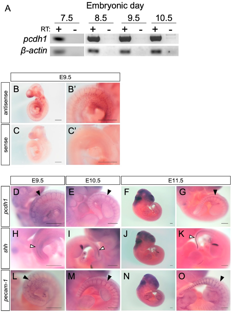
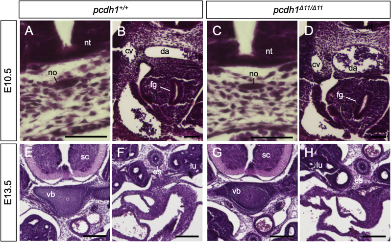
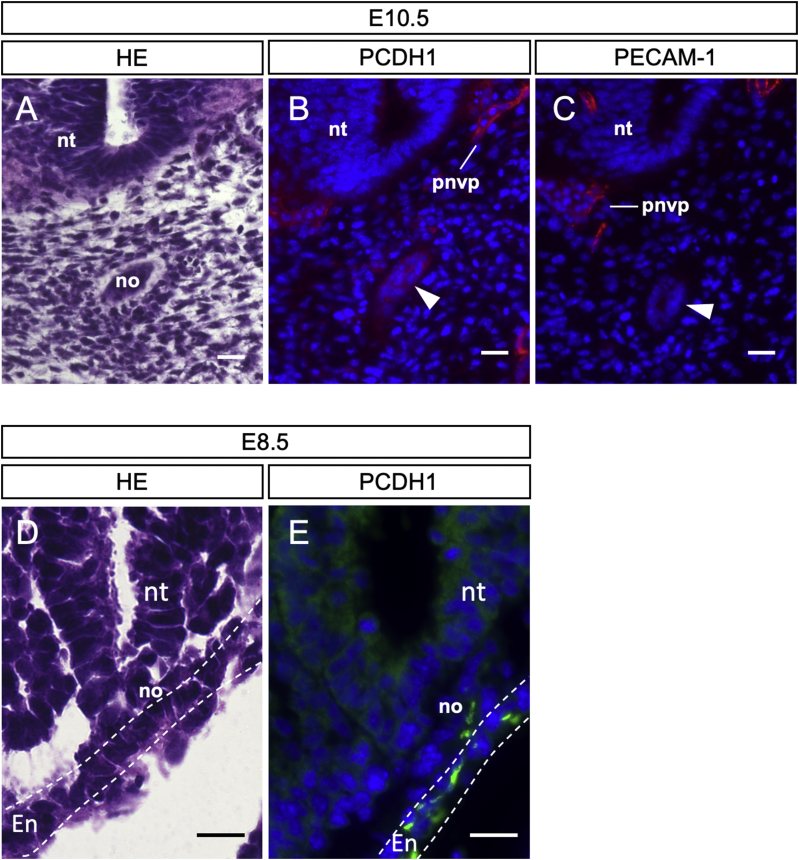
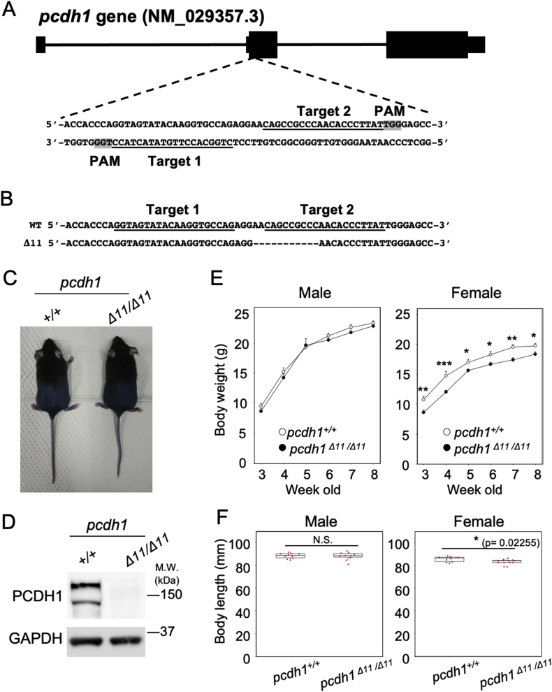
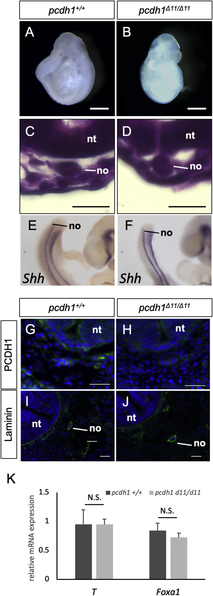
Notochord is an embryonic midline structure that serves as mechanical support for axis elongation and the signaling center for the surrounding tissues. Precursors of notochord are initially induced in the dorsal most mesoderm region in gastrulating embryo and separate from the surrounding mesoderm/endoderm tissue to form an elongated rod-like structure, suggesting that cell adhesion molecules may play an important role in this step. In embryo, axial protocadherin (AXPC), an orthologue of mammalian Protocadherin-1 (PCDH1), is indispensable for the assembly and separation from the surrounding tissue of the notochord cells. However, the role of PCDH1 in mammalian notochord remains unknown. We herein report that PCDH1 is expressed in the notochord of mouse embryo and that PCDH1-deficient mice form notochord normally. First, we examined the temporal expression pattern of and found that mRNA was expressed from embryonic day (E) 7.5, prior to the stage when notochord cells detach from the surrounding endoderm tissue. Second, we found that PCDH1 protein is expressed in the notochord of mouse embryos in addition to the previously reported expression in endothelial cells. To further investigate the role of PCDH1 in embryonic development, we generated PCDH1-deficient mice using the CRISPR-Cas9 system. In PCDH1-deficient embryos, notochord formation and separation from the surrounding tissue were normal. Structure and marker gene expression of notochord were also unaffected by loss of PCDH1. Major vascular patterns in PCDH1-deficient embryo were essentially normal. These results suggest that PCDH1 is dispensable for notochord formation, including the tissue separation process, in mammalian embryos. We successfully identified the evolutionary conserved expression of PCDH1 in notochord, but its function may differ among species.
脊索是一种胚胎中线结构,为轴伸长提供机械支持,并作为周围组织的信号中心。脊索的前体最初在原肠胚形成期胚胎的最背侧中胚层区域被诱导产生,并与周围的中胚层/内胚层组织分离,形成一个细长的杆状结构,这表明细胞粘附分子可能在这一步骤中发挥重要作用。在胚胎中,轴向原钙粘蛋白(AXPC)是哺乳动物原钙粘蛋白-1(PCDH1)的同源物,对于脊索细胞的组装以及与周围组织的分离是必不可少的。然而,PCDH1在哺乳动物脊索中的作用仍然未知。我们在此报告,PCDH1在小鼠胚胎的脊索中表达,并且PCDH1基因敲除小鼠的脊索形成正常。首先,我们检查了其时间表达模式,发现PCDH1 mRNA在胚胎第7.5天(E7.5)就开始表达,早于脊索细胞与周围内胚层组织分离的阶段。其次,我们发现PCDH1蛋白除了先前报道的在内皮细胞中的表达外,还在小鼠胚胎的脊索中表达。为了进一步研究PCDH1在胚胎发育中的作用,我们使用CRISPR-Cas9系统生成了PCDH1基因敲除小鼠。在PCDH1基因敲除胚胎中,脊索的形成以及与周围组织的分离是正常的。脊索的结构和标记基因表达也不受PCDH1缺失的影响。PCDH1基因敲除胚胎中的主要血管模式基本正常。这些结果表明,PCDH1对于哺乳动物胚胎中的脊索形成,包括组织分离过程,是可有可无的。我们成功鉴定了PCDH1在脊索中的进化保守表达,但其功能可能在不同物种间存在差异。