• 文献检索
  • 文档翻译
  • 深度研究
  • 学术资讯
  • Suppr Zotero 插件Zotero 插件
  • 邀请有礼
  • 套餐&价格
  • 历史记录
应用&插件
Suppr Zotero 插件Zotero 插件浏览器插件Mac 客户端Windows 客户端微信小程序
定价
高级版会员购买积分包购买API积分包
服务
文献检索文档翻译深度研究API 文档MCP 服务
关于我们
关于 Suppr公司介绍联系我们用户协议隐私条款
关注我们

Suppr 超能文献

核心技术专利:CN118964589B侵权必究
粤ICP备2023148730 号-1Suppr @ 2026

文献检索

告别复杂PubMed语法,用中文像聊天一样搜索,搜遍4000万医学文献。AI智能推荐,让科研检索更轻松。

立即免费搜索

文件翻译

保留排版,准确专业,支持PDF/Word/PPT等文件格式,支持 12+语言互译。

免费翻译文档

深度研究

AI帮你快速写综述,25分钟生成高质量综述,智能提取关键信息,辅助科研写作。

立即免费体验

组织光学透明化的发展能否辅助超分辨率显微镜成像?

Can Developments in Tissue Optical Clearing Aid Super-Resolution Microscopy Imaging?

机构信息

Department of Immunology, Medical University of Warsaw, 02-097 Warsaw, Poland.

The Doctoral School of the Medical University of Warsaw, Medical University of Warsaw, 02-097 Warsaw, Poland.

出版信息

Int J Mol Sci. 2021 Jun 23;22(13):6730. doi: 10.3390/ijms22136730.

DOI:10.3390/ijms22136730
PMID:34201632
原文链接:https://pmc.ncbi.nlm.nih.gov/articles/PMC8268743/
Abstract

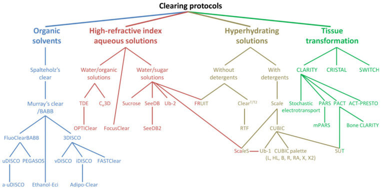
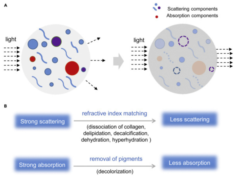
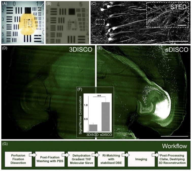
The rapid development of super-resolution microscopy (SRM) techniques opens new avenues to examine cell and tissue details at a nanometer scale. Due to compatibility with specific labelling approaches, in vivo imaging and the relative ease of sample preparation, SRM appears to be a valuable alternative to laborious electron microscopy techniques. SRM, however, is not free from drawbacks, with the rapid quenching of the fluorescence signal, sensitivity to spherical aberrations and light scattering that typically limits imaging depth up to few micrometers being the most pronounced ones. Recently presented and robustly optimized sets of tissue optical clearing (TOC) techniques turn biological specimens transparent, which greatly increases the tissue thickness that is available for imaging without loss of resolution. Hence, SRM and TOC are naturally synergistic techniques, and a proper combination of these might promptly reveal the three-dimensional structure of entire organs with nanometer resolution. As such, an effort to introduce large-scale volumetric SRM has already started; in this review, we discuss TOC approaches that might be favorable during the preparation of SRM samples. Thus, special emphasis is put on TOC methods that enhance the preservation of fluorescence intensity, offer the homogenous distribution of molecular probes, and vastly decrease spherical aberrations. Finally, we review examples of studies in which both SRM and TOC were successfully applied to study biological systems.

摘要

超分辨率显微镜 (SRM) 技术的快速发展为在纳米尺度上检查细胞和组织细节开辟了新途径。由于与特定标记方法的兼容性、体内成像以及相对容易的样品制备,SRM 似乎是一种替代繁琐的电子显微镜技术的有价值的方法。然而,SRM 并非没有缺点,最明显的缺点是荧光信号的快速猝灭、对球差和光散射的敏感性,这些通常限制了成像深度至几微米。最近提出的和经过稳健优化的一系列组织光学透明化 (TOC) 技术使生物样本变得透明,这大大增加了可用于成像而不会损失分辨率的组织厚度。因此,SRM 和 TOC 是自然协同的技术,这些技术的适当结合可能会立即以纳米分辨率揭示整个器官的三维结构。因此,已经开始努力引入大规模体积 SRM;在这篇综述中,我们讨论了在 SRM 样品制备过程中可能有利的 TOC 方法。因此,特别强调了那些增强荧光强度保持、提供分子探针均匀分布以及大大降低球差的 TOC 方法。最后,我们回顾了成功应用于研究生物系统的 SRM 和 TOC 的例子。

https://cdn.ncbi.nlm.nih.gov/pmc/blobs/0ccf/8268743/1179d8cc82f6/ijms-22-06730-g007.jpg
https://cdn.ncbi.nlm.nih.gov/pmc/blobs/0ccf/8268743/f80c18c7ee0e/ijms-22-06730-g001.jpg
https://cdn.ncbi.nlm.nih.gov/pmc/blobs/0ccf/8268743/e586e24d6908/ijms-22-06730-g002.jpg
https://cdn.ncbi.nlm.nih.gov/pmc/blobs/0ccf/8268743/22315f0af48b/ijms-22-06730-g003.jpg
https://cdn.ncbi.nlm.nih.gov/pmc/blobs/0ccf/8268743/012b5407e281/ijms-22-06730-g004.jpg
https://cdn.ncbi.nlm.nih.gov/pmc/blobs/0ccf/8268743/3bb5aaf208ed/ijms-22-06730-g005.jpg
https://cdn.ncbi.nlm.nih.gov/pmc/blobs/0ccf/8268743/9cf913b2c4fe/ijms-22-06730-g006.jpg
https://cdn.ncbi.nlm.nih.gov/pmc/blobs/0ccf/8268743/1179d8cc82f6/ijms-22-06730-g007.jpg

相似文献

1
Can Developments in Tissue Optical Clearing Aid Super-Resolution Microscopy Imaging?组织光学透明化的发展能否辅助超分辨率显微镜成像?
Int J Mol Sci. 2021 Jun 23;22(13):6730. doi: 10.3390/ijms22136730.
2
Imaging the mammary gland and mammary tumours in 3D: optical tissue clearing and immunofluorescence methods.三维成像乳腺及乳腺肿瘤:光学组织透明化和免疫荧光方法
Breast Cancer Res. 2016 Dec 13;18(1):127. doi: 10.1186/s13058-016-0754-9.
3
A method for ultrafast tissue clearing that preserves fluorescence for multimodal and longitudinal brain imaging.一种超快组织透明化方法,可保持荧光用于多模态和纵向脑成像。
BMC Biol. 2022 Mar 29;20(1):77. doi: 10.1186/s12915-022-01275-6.
4
Super-resolution microscopy: a brief history and new avenues.超分辨率显微镜:简史与新途径。
Philos Trans A Math Phys Eng Sci. 2022 Apr 4;380(2220):20210110. doi: 10.1098/rsta.2021.0110. Epub 2022 Feb 14.
5
Clarity and Immunofluorescence on Mouse Brain Tissue.小鼠脑组织的清晰度和免疫荧光
Curr Protoc Neurosci. 2018 Apr;83(1):e46. doi: 10.1002/cpns.46.
6
UbasM: An effective balanced optical clearing method for intact biomedical imaging.UbasM:一种用于完整生物医学成像的有效平衡光透明方法。
Sci Rep. 2017 Sep 22;7(1):12218. doi: 10.1038/s41598-017-12484-3.
7
Fluorescence fluctuation-based super-resolution microscopy: Basic concepts for an easy start.基于荧光波动的超分辨率显微镜:易于入门的基本概念。
J Microsc. 2022 Dec;288(3):218-241. doi: 10.1111/jmi.13135. Epub 2022 Aug 9.
8
Super-resolution Microscopy for Biological Imaging.超分辨率显微镜在生物成像中的应用。
Adv Exp Med Biol. 2021;3233:23-43. doi: 10.1007/978-981-15-7627-0_2.
9
Tissue clearing for optical anatomy.组织透明化用于光学解剖。
Angew Chem Int Ed Engl. 2013 Oct 11;52(42):10949-51. doi: 10.1002/anie.201306039. Epub 2013 Sep 3.
10
Functional imaging in bulk tissue specimens using optical emission tomography: fluorescence preservation during optical clearing.使用光学发射断层扫描对大块组织标本进行功能成像:光学透明过程中的荧光保存。
Phys Med Biol. 2007 Apr 21;52(8):2035-54. doi: 10.1088/0031-9155/52/8/001. Epub 2007 Mar 20.

本文引用的文献

1
Rapid methods for the evaluation of fluorescent reporters in tissue clearing and the segmentation of large vascular structures.用于在组织透明化中评估荧光报告基因及对大型血管结构进行分割的快速方法。
iScience. 2021 May 26;24(6):102650. doi: 10.1016/j.isci.2021.102650. eCollection 2021 Jun 25.
2
Physical and chemical mechanisms of tissue optical clearing.组织光学透明化的物理和化学机制。
iScience. 2021 Feb 12;24(3):102178. doi: 10.1016/j.isci.2021.102178. eCollection 2021 Mar 19.
3
Efficient Tissue Clearing and Multi-Organ Volumetric Imaging Enable Quantitative Visualization of Sparse Immune Cell Populations During Inflammation.
高效的组织通透化和多器官容积成像可实现炎症过程中稀疏免疫细胞群体的定量可视化。
Front Immunol. 2021 Jan 25;11:599495. doi: 10.3389/fimmu.2020.599495. eCollection 2020.
4
Comparison of Transparency and Shrinkage During Clearing of Insect Brains Using Media With Tunable Refractive Index.使用具有可调折射率的介质对昆虫大脑进行透明化处理期间的透明度和收缩率比较
Front Neuroanat. 2020 Nov 20;14:599282. doi: 10.3389/fnana.2020.599282. eCollection 2020.
5
mGreenLantern: a bright monomeric fluorescent protein with rapid expression and cell filling properties for neuronal imaging.mGreenLantern:一种明亮的单体荧光蛋白,具有快速表达和细胞填充特性,可用于神经元成像。
Proc Natl Acad Sci U S A. 2020 Dec 1;117(48):30710-30721. doi: 10.1073/pnas.2000942117. Epub 2020 Nov 18.
6
Rapid immunostaining method for three-dimensional volume imaging of biological tissues by magnetic force-induced focusing of the electric field.利用磁场诱导的电场聚焦快速免疫染色方法对生物组织进行三维体积成像。
Brain Struct Funct. 2021 Jan;226(1):297-309. doi: 10.1007/s00429-020-02160-0. Epub 2020 Nov 11.
7
A Versatile Tiling Light Sheet Microscope for Imaging of Cleared Tissues.一种通用的平铺光片显微镜,用于对透明组织进行成像。
Cell Rep. 2020 Nov 3;33(5):108349. doi: 10.1016/j.celrep.2020.108349.
8
Super-resolution Microscopy with Single Molecules in Biology and Beyond-Essentials, Current Trends, and Future Challenges.超越生物学的单分子超分辨率显微镜——要点、当前趋势和未来挑战。
J Am Chem Soc. 2020 Oct 21;142(42):17828-17844. doi: 10.1021/jacs.0c08178. Epub 2020 Oct 9.
9
Biomedical Applications of Tissue Clearing and Three-Dimensional Imaging in Health and Disease.组织透明化和三维成像在健康与疾病中的生物医学应用
iScience. 2020 Aug 21;23(8):101432. doi: 10.1016/j.isci.2020.101432. Epub 2020 Aug 2.
10
Molecular resolution imaging by post-labeling expansion single-molecule localization microscopy (Ex-SMLM).通过标记后扩展单分子定位显微镜(Ex-SMLM)进行分子分辨率成像。
Nat Commun. 2020 Jul 7;11(1):3388. doi: 10.1038/s41467-020-17086-8.