Thompson Atalie C, Li Ang, Asrani Sanjay
Department of Ophthalmology, Duke University, Box 3802, Durham, NC, 27710, USA.
Cleveland Clinic, Cleveland, OH, USA.
Ophthalmol Ther. 2021 Sep;10(3):629-642. doi: 10.1007/s40123-021-00355-0. Epub 2021 Jul 1.
To evaluate the agreement between trend-based analysis and qualitative assessment of the retinal nerve fiber layer (RNFL) thickness for glaucomatous progression on spectral-domain optical coherence tomography (SDOCT).
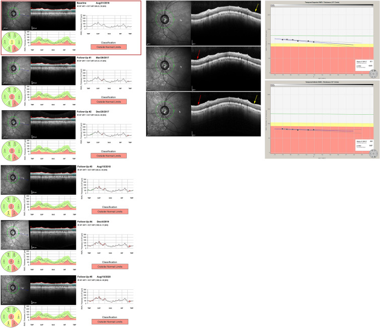
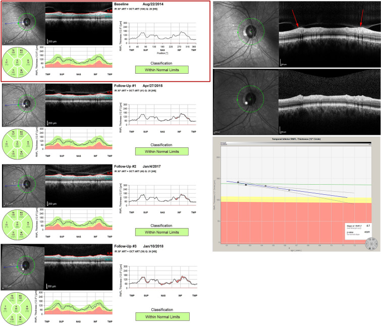
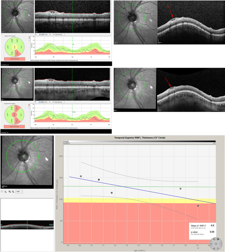
Retrospective review of 190 eyes from 103 patients with glaucoma or suspected glaucoma that underwent SDOCT imaging during four consecutive clinic visits. Trend-based progression was characterized by a significantly negative slope. Progression by qualitative analysis was determined by review of raw SDOCT B-scans.
The slope was significantly greater in those with progression than without progression for both trend-based and qualitative analysis (p < 0.001). However, the qualitative grading classified a significantly greater proportion of eyes as progressing compared to trend-based analysis in both the superotemporal (ST) (23.2% vs. 10.5%, p = 0.001) and inferotemporal (IT) RNFL (27.4% vs 8.4%, p < 0.001). The trend-based and qualitative classifications of progression showed poor agreement in both the ST (kappa = 0.0135) and IT RNFL (kappa = 0.1222). The agreement between trend-based and qualitative analysis was lower for eyes with artifacts (ST = 58.11%; IT = 68.7%) than those without artifacts (ST = 80.2%; IT = 74.8%). Moreover, among eyes with artifacts, there was no significant difference in slope between those qualitatively categorized as progressing versus not progressing (p > 0.05).
Poor agreement was found between a trend-based and qualitative analysis of change in RNFL on SDOCT. Careful qualitative review of SDOCT imaging may identify specific areas of glaucoma progression not captured by trend-based methods, especially in the presence of artifacts. Such an approach may also prove useful for detecting glaucoma progression in a clinical setting when there are few data points available.
评估基于趋势分析与视网膜神经纤维层(RNFL)厚度的定性评估在光谱域光学相干断层扫描(SDOCT)上对青光眼进展的一致性。
回顾性分析103例青光眼或疑似青光眼患者的190只眼睛,这些眼睛在连续四次门诊就诊期间接受了SDOCT成像。基于趋势的进展以显著的负斜率为特征。通过回顾原始SDOCT B扫描来确定定性分析的进展情况。
对于基于趋势和定性分析,进展者的斜率均显著大于无进展者(p < 0.001)。然而,与基于趋势的分析相比,定性分级在颞上(ST)(23.2%对10.5%,p = 0.001)和颞下(IT)RNFL(27.4%对8.4%,p < 0.001)将更多比例的眼睛分类为进展。基于趋势和定性的进展分类在ST(kappa = 0.0135)和IT RNFL(kappa = 0.1222)中一致性较差。有伪像的眼睛中基于趋势和定性分析的一致性低于无伪像的眼睛(ST = 58.11%;IT = 68.7%)与无伪像的眼睛(ST = 80.2%;IT = 74.8%)。此外,在有伪像的眼睛中,定性分类为进展与未进展的眼睛之间斜率无显著差异(p > 0.05)。
发现基于趋势的分析与SDOCT上RNFL变化的定性分析之间一致性较差。对SDOCT成像进行仔细的定性回顾可能会识别出基于趋势的方法未捕捉到的青光眼进展的特定区域,尤其是在存在伪像的情况下。当可用数据点较少时,这种方法在临床环境中检测青光眼进展方面可能也很有用。