Department of Orthopedic Surgery, Kindai University Hospital, Osaka-Sayama City, Osaka.
Eur J Histochem. 2021 Jul 2;65(3):3203. doi: 10.4081/ejh.2021.3203.
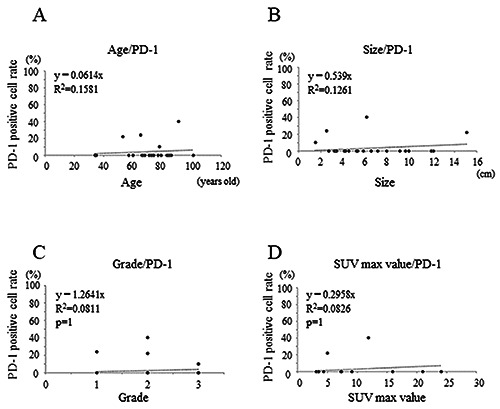
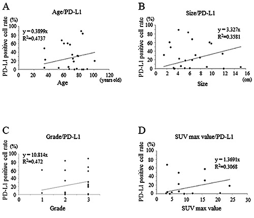
Inhibitors of the programmed death-1/programmed death-ligand 1 (PD-1/PD-L1) immune checkpoint system are used for treating various malignancies. However, evidence on their use in soft tissue sarcomas (STS) is limited. This study aimed to retrospectively investigate the relationship between the expression of PD-1/PD-L1 and related antigens in STS, and their association with clinical characteristics. Immunostaining for CD4, CD8, PD-1, PD-L1, IL-2, and IFN-γ was performed using pathological specimens harvested at the time of biopsy from 10 patients with undifferentiated pleomorphic sarcoma (UPS), nine with myxofibrosarcoma (MFS), and three with malignant peripheral nerve sheath tumor (MPNST) who were treated at our hospital. Subsequently, the positive immunostaining cell rates were calculated. We also examined the correlation between each immune positive cell rate and age, tissue grade, size, and maximum standardized uptake (SUV-max) values. The 3-year event-free survival (EFS) and overall survival (OS) rates were compared between the positive and negative groups (positive rate >10%; negative <10%) for various immune stains. The positive rates were also compared between the presence and absence of events groups. There was positive staining for the immune checkpoint molecules in every STS type except for PD-1 in MPNST. CD4, CD8, and PD-1 stained lymphocytes in close proximity to the tumor in adjacent tissue sections. A positive correlation was observed between the positive cell rates of each immune component including inflammatory cytokines such as IL-2 and IFN-γ. Additionally, the clinical features positively correlated with the positive PD-1/PD-L1 expression rates. No significant differences in the 3-EFS and OS rates was observed between the PD-1/PD-L1 positive and negative groups. Our results suggest that an inducible immune checkpoint mechanism may be involved in UPS, MFS, and MPNST.
程序性死亡受体-1/程序性死亡配体 1(PD-1/PD-L1)免疫检查点抑制剂被用于治疗各种恶性肿瘤。然而,关于其在软组织肉瘤(STS)中的应用证据有限。本研究旨在回顾性研究 STS 中 PD-1/PD-L1 及其相关抗原的表达与临床特征的关系。对在我院治疗的 10 例未分化多形性肉瘤(UPS)、9 例黏液纤维肉瘤(MFS)和 3 例恶性外周神经鞘瘤(MPNST)患者的活检标本进行了 CD4、CD8、PD-1、PD-L1、IL-2 和 IFN-γ 的免疫组织化学染色,随后计算了阳性免疫染色细胞率。我们还检查了每种免疫阳性细胞率与年龄、组织分级、大小和最大标准化摄取值(SUV-max)值之间的相关性。比较了各种免疫染色阳性和阴性组(阳性率>10%;阴性率<10%)之间的 3 年无事件生存(EFS)和总生存(OS)率。还比较了有事件和无事件组之间的阳性率。除 MPNST 外,每种 STS 类型均存在免疫检查点分子的阳性染色。CD4、CD8 和 PD-1 在相邻组织切片中染色靠近肿瘤的淋巴细胞。包括炎性细胞因子如 IL-2 和 IFN-γ 在内的每种免疫成分的阳性细胞率呈正相关。此外,临床特征与 PD-1/PD-L1 表达率呈正相关。PD-1/PD-L1 阳性和阴性组之间的 3 年 EFS 和 OS 率没有显著差异。我们的结果表明,诱导性免疫检查点机制可能参与 UPS、MFS 和 MPNST 的发生。