Suppr超能文献

一种应用于人类牙釉质的亚微米级晶体学有序/无序映射方法。

A method for mapping submicron-scale crystallographic order/disorder applied to human tooth enamel.

作者信息

Free R, DeRocher K, Xu R, Joester D, Stock S R

机构信息

Department of Materials Science and Engineering, Northwestern University, Evanston, Illinois, USA.

Argonne National Lab, Advanced Photon Source, Lemont, Illinois 34ID-E, USA.

出版信息

Powder Diffr. 2020 Jun;35(2):117-123. doi: 10.1017/s0885715620000251. Epub 2020 May 8.

Abstract

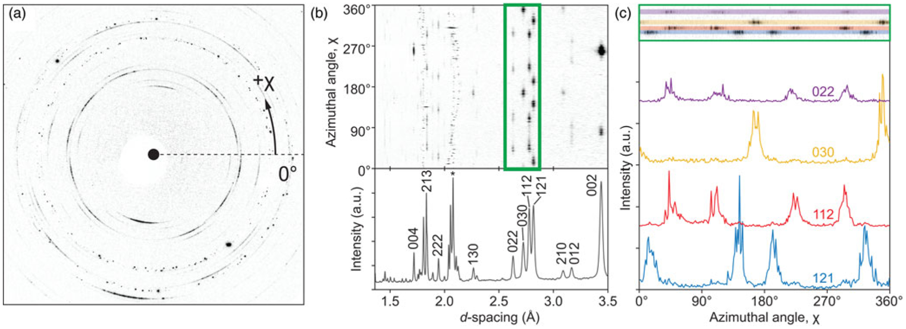
Tooth enamel, the outermost layer of human teeth, is a complex, hierarchically structured biocomposite. The details of this structure are important in multiple human health contexts, from understanding the progression of dental caries (tooth decay) to understanding the process of amelogenesis and related developmental defects. Enamel is composed primarily of long, nanoscale crystallites of hydroxyapatite that are bundled by the thousands to form micron-scale rods. Studies with transmission electron microscopy show the relationships between small groups of crystallites and X-ray diffraction characterize averages over many rods, but the direct measurement of variations in local crystallographic structure across and between enamel rods has been missing. Here, we describe a synchrotron X-ray-based experimental approach and a novel analysis method developed to address this gap in knowledge. A 500-nm-wide beam of monochromatic X-rays in conjunction with a sample section only 1 μm in thickness enables 2D diffraction patterns to be collected from small well-separated volumes within the enamel microstructure but still probes enough crystallites (300 per pattern) to extract population-level statistics on crystallographic features like lattice parameter, crystallite size, and orientation distributions. Furthermore, the development of a quantitative metric to characterize relative order and disorder based on the azimuthal autocorrelation of diffracted intensity enables these crystallographic measurements to be correlated with their location within the enamel microstructure (e.g., between rod and interrod regions). These methods represent a step forward in the characterization of human enamel and will elucidate the variation of the crystallographic structure across and between enamel rods for the first time.

摘要

牙釉质是人类牙齿的最外层,是一种复杂的、具有层次结构的生物复合材料。这种结构的细节在多种人类健康背景下都很重要,从理解龋齿(蛀牙)的发展到理解釉质形成过程及相关发育缺陷。牙釉质主要由长的纳米级羟基磷灰石微晶组成,这些微晶数以千计地聚集在一起形成微米级的棒状物。透射电子显微镜研究显示了小群微晶之间的关系,X射线衍射则表征了许多棒状物的平均值,但牙釉质棒之间和内部局部晶体结构变化的直接测量一直缺失。在此,我们描述了一种基于同步加速器X射线的实验方法和一种新开发的分析方法,以填补这一知识空白。一束约500纳米宽的单色X射线与仅1微米厚的样品切片相结合,能够从牙釉质微观结构中分离良好的小体积区域收集二维衍射图案,但仍能探测到足够数量的微晶(每个图案约300个),以提取诸如晶格参数、微晶尺寸和取向分布等晶体学特征的总体统计数据。此外,基于衍射强度方位自相关开发的一种定量指标来表征相对有序和无序,使得这些晶体学测量能够与它们在牙釉质微观结构中的位置相关联(例如,在棒状区域和棒间区域之间)。这些方法代表了人类牙釉质表征方面的一个进步,并将首次阐明牙釉质棒之间和内部晶体结构的变化。

相似文献

1
A method for mapping submicron-scale crystallographic order/disorder applied to human tooth enamel.
Powder Diffr. 2020 Jun;35(2):117-123. doi: 10.1017/s0885715620000251. Epub 2020 May 8.
2
Mesoscale structural gradients in human tooth enamel.
Proc Natl Acad Sci U S A. 2022 Dec 27;119(52):e2211285119. doi: 10.1073/pnas.2211285119. Epub 2022 Dec 19.
3
In situ monitoring and analysis of enamel demineralisation using synchrotron X-ray scattering.
Acta Biomater. 2018 Sep 1;77:333-341. doi: 10.1016/j.actbio.2018.07.027. Epub 2018 Jul 17.
4
Enamelin Directs Crystallite Organization at the Enamel-Dentine Junction.
J Dent Res. 2016 May;95(5):580-7. doi: 10.1177/0022034516632745. Epub 2016 Feb 24.
5
Mapping the spatial and temporal progression of human dental enamel biomineralization using synchrotron X-ray diffraction.
Arch Oral Biol. 2013 Nov;58(11):1726-34. doi: 10.1016/j.archoralbio.2013.08.012. Epub 2013 Aug 31.
8
2D mapping of texture and lattice parameters of dental enamel.
Biomaterials. 2007 Jun;28(18):2908-14. doi: 10.1016/j.biomaterials.2007.02.019. Epub 2007 Feb 25.
9
Intracoronal stress transfer through enamel following RBC photopolymerisation: A synchrotron X-ray study.
Dent Mater. 2018 Oct;34(10):1426-1439. doi: 10.1016/j.dental.2018.07.005. Epub 2018 Aug 14.

引用本文的文献

1
Stratification of fluoride uptake among enamel crystals with age elucidated by atom probe tomography.
Commun Mater. 2024;5(1):270. doi: 10.1038/s43246-024-00709-8. Epub 2024 Dec 19.
2
Adult dental epithelial stem cell-derived organoids deposit hydroxylapatite biomineral.
Int J Oral Sci. 2023 Dec 7;15(1):55. doi: 10.1038/s41368-023-00257-w.
3
Multi-resolution Correlative Ultrastructural and Chemical Analysis of Carious Enamel by Scanning Microscopy and Tomographic Imaging.
ACS Appl Mater Interfaces. 2023 Aug 9;15(31):37259-37273. doi: 10.1021/acsami.3c08031. Epub 2023 Jul 31.
5
Mesoscale structural gradients in human tooth enamel.
Proc Natl Acad Sci U S A. 2022 Dec 27;119(52):e2211285119. doi: 10.1073/pnas.2211285119. Epub 2022 Dec 19.
6
Bioapatite in shark centra studied by wide-angle and by small-angle X-ray scattering.
J R Soc Interface. 2022 Sep;19(194):20220373. doi: 10.1098/rsif.2022.0373. Epub 2022 Sep 21.
7
Combination of Two Synchrotron Radiation-Based Techniques and Chemometrics to Study an Enhanced Natural Remineralization of Enamel.
Anal Chem. 2022 Apr 5;94(13):5359-5366. doi: 10.1021/acs.analchem.1c05498. Epub 2022 Mar 23.
8
Microstructure and energy dispersive diffraction reconstruction of 3D patterns of crystallographic texture in a shark centrum.
J Med Imaging (Bellingham). 2022 May;9(3):031504. doi: 10.1117/1.JMI.9.3.031504. Epub 2022 Feb 2.

本文引用的文献

2
Materials engineering by ameloblasts.
J Dent Res. 2015 Jun;94(6):759-67. doi: 10.1177/0022034515577963. Epub 2015 Mar 23.
3
Dental materials. Amorphous intergranular phases control the properties of rodent tooth enamel.
Science. 2015 Feb 13;347(6223):746-50. doi: 10.1126/science.1258950.
4
X-ray microdiffraction, TEM characterization and texture analysis of human dentin and enamel.
J Microsc. 2013 Aug;251(2):144-53. doi: 10.1111/jmi.12053. Epub 2013 May 29.
5
Disruption of enamel crystal formation quantified by synchrotron microdiffraction.
J Dent. 2012 Dec;40(12):1074-80. doi: 10.1016/j.jdent.2012.08.020. Epub 2012 Sep 7.
6
NIH Image to ImageJ: 25 years of image analysis.
Nat Methods. 2012 Jul;9(7):671-5. doi: 10.1038/nmeth.2089.
7
Distribution of enamel crystallite orientation through an entire tooth crown studied using synchrotron X-ray diffraction.
Eur J Oral Sci. 2011 Dec;119 Suppl 1:19-24. doi: 10.1111/j.1600-0722.2011.00909.x.
8
2D mapping of texture and lattice parameters of dental enamel.
Biomaterials. 2007 Jun;28(18):2908-14. doi: 10.1016/j.biomaterials.2007.02.019. Epub 2007 Feb 25.
9
Enamel - a "metallic-like" deformable biocomposite.
J Dent. 2007 May;35(5):431-7. doi: 10.1016/j.jdent.2006.12.002. Epub 2007 Jan 31.
10
High-resolution electron microscopy of enamel-crystal demineralization and remineralization in carious lesions.
J Electron Microsc (Tokyo). 2003;52(6):605-13. doi: 10.1093/jmicro/52.6.605.

文献AI研究员

20分钟写一篇综述,助力文献阅读效率提升50倍。

立即体验

用中文搜PubMed

大模型驱动的PubMed中文搜索引擎

马上搜索

文档翻译

学术文献翻译模型,支持多种主流文档格式。

立即体验