Department of Pediatrics, Faculty of Medicine, Cairo University, Cairo, Egypt.
Department of Radiodiagnosis, Faculty of Medicine, Cairo University, Cairo, Egypt.
F1000Res. 2020 Sep 9;9:1108. doi: 10.12688/f1000research.25943.2. eCollection 2020.
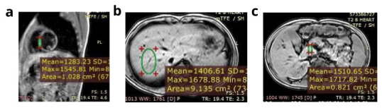
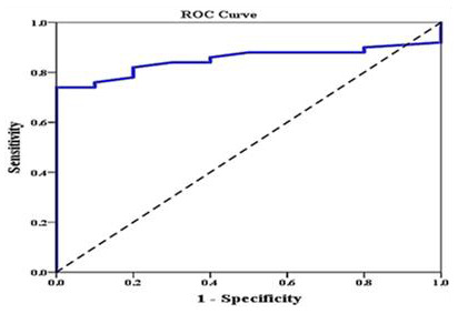
Cardiac, hepatic and pancreatic T2* measured by magnetic resonance imaging (MRI) has been proven to be an accurate and non-invasive method for measuring iron overload in iron overload conditions. There is accumulating evidence that pancreatic iron can predict cardiac iron in young children because the pancreas loads earlier than the heart. The aim of our study was to assess the relationships between pancreatic T2* values and pancreatic iron loading with cardiac dysfunctions and liver and cardiac iron among patients with β-thalassaemia major (βTM) and sickle cell disease (SCD). 40 βTM and 20 transfusion-dependant SCD patients were included along with 60 healthy age and sex-matched controls. Echocardiography and Tissue Doppler Imaging were performed for all subjects as well as the control group. Hepatic, cardiac and pancreatic iron overload in cases were assessed by MRI T2*. The mean age of our patients was 13.7 years with mean frequency of transfusion/year 12. Mean cardiac T2* was 32.9 ms and mean myocardial iron concentration was 0.7 mg/g; One patient had cardiac iron overload of moderate severity. Mean pancreatic T2* was 22.3 ms with 20 patients having mild pancreatic iron overload. Pancreatic T2* correlated positively peak late diastolic velocity at septal mitral annulus (r=0.269, p=0.038), peak early diastolic velocity at tricuspid annulus (r=0.430, p=0.001) and mitral annular plane systolic excursion (r=0.326, p=0.01); and negatively with end systolic pulmonary artery pressure (r=-0.343, p=0.007) and main pulmonary artery diameter (MPA) (r=-0.259, p=0.046). We couldn't test the predictability of pancreatic T2* in relation to cardiac T2* as only one patient had cardiac T2*<20 ms. : There was a relationship between pancreatic iron siderosis with cardiac dysfunction in multi-transfused patients with βTM and SCD. No direct relation between pancreatic iron and cardiac siderosis was detected.
心脏、肝脏和胰腺的 T2* 通过磁共振成像(MRI)测量,已被证明是测量铁过载的准确和非侵入性方法。越来越多的证据表明,胰腺铁可以预测幼儿的心脏铁,因为胰腺比心脏更早负荷。我们的研究目的是评估 40 名β地中海贫血(βTM)和镰状细胞病(SCD)患者的胰腺 T2* 值与胰腺铁负荷与心脏功能障碍以及肝脏和心脏铁之间的关系。 还包括 20 名输血依赖的 SCD 患者和 60 名年龄和性别匹配的健康对照组。所有受试者以及对照组均进行了超声心动图和组织多普勒成像。 通过 MRI T2* 评估病例中的肝脏、心脏和胰腺铁过载。 我们患者的平均年龄为 13.7 岁,平均每年输血 12 次。平均心脏 T2为 32.9ms,平均心肌铁浓度为 0.7mg/g;有 1 名患者心脏铁过载程度为中度。平均胰腺 T2为 22.3ms,20 名患者轻度胰腺铁过载。胰腺 T2与二尖瓣环间隔晚期舒张峰值速度呈正相关(r=0.269,p=0.038),三尖瓣环早期舒张峰值速度呈正相关(r=0.430,p=0.001)和二尖瓣环平面收缩期位移(r=0.326,p=0.01);与收缩末期肺动脉压(r=-0.343,p=0.007)和主肺动脉直径(MPA)(r=-0.259,p=0.046)呈负相关。我们不能测试胰腺 T2与心脏 T2之间的预测关系,因为只有 1 名患者的心脏 T2<20ms。:在接受多次输血的βTM 和 SCD 患者中,胰腺铁沉积与心脏功能障碍之间存在关系。未检测到胰腺铁与心脏铁沉积之间的直接关系。