Department of Neurology, Zhujiang Hospital, Southern Medical University, Guangzhou, China.
Department of Neurology, Second School of Clinical Medicine, Southern Medical University, Guangzhou, China.
Bioengineered. 2021 Dec;12(1):3957-3967. doi: 10.1080/21655979.2021.1947630.
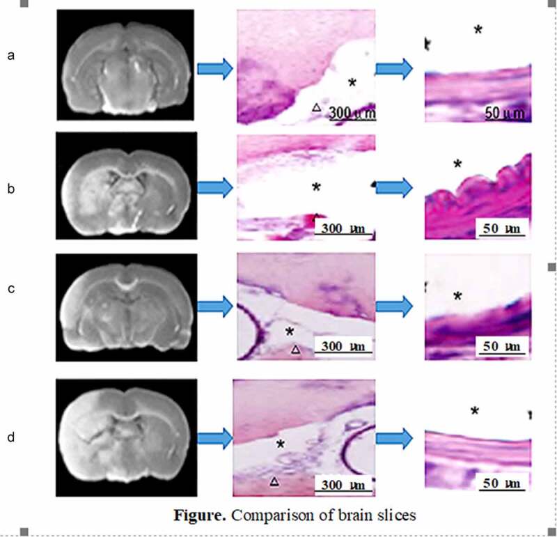
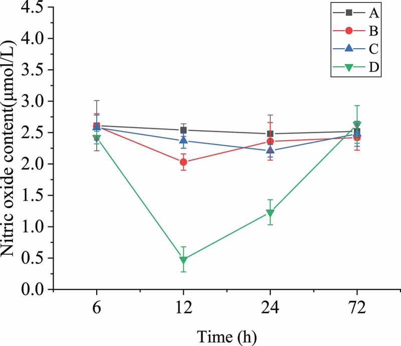
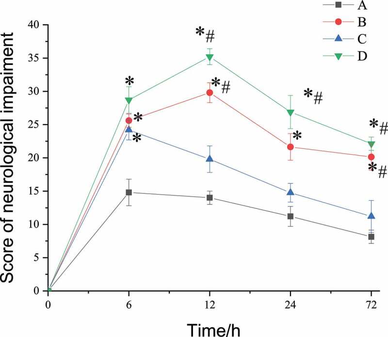
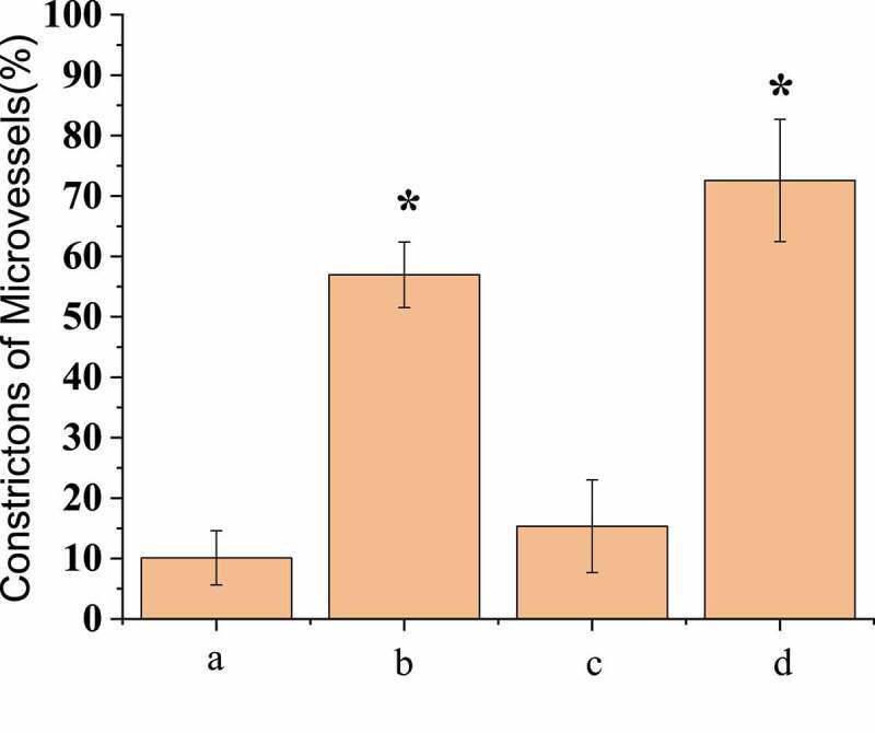
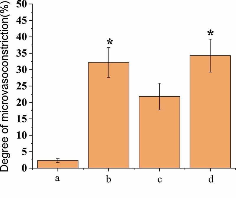
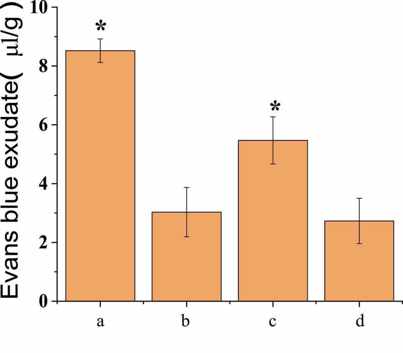
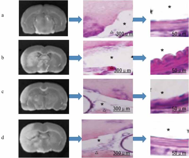
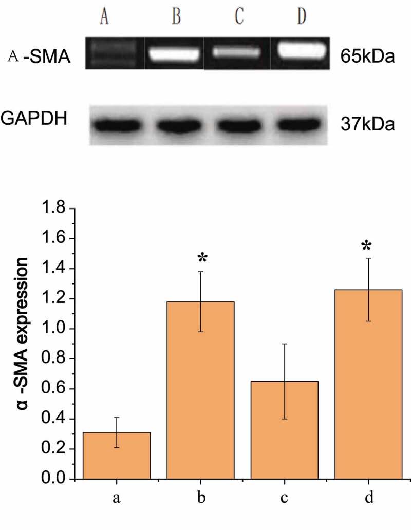
To investigate mechanism of pericytes in the early stage of subarachnoid haemorrhage (SAH) and its associated microvascular spasm and neurovascular injury, 100 healthy 8-week-old Sprague-Dawley male rats were taken as subjects and divided into four groups: group A (sham operation, control group), group B (SAH operation group), group C (SAH operation group treated with scutellarin), and group D (SAH operation group treated with L-nitro-arginine). 72 hours after the operation, the rats were conducted assessment of neurological impairment, observation of microangiography, detection of blood-brain barrier permeability, observation of skull base haemorrhage, identification of pericyte culture, and measurement of blood nitric oxide. The results showed that neurological impairment score, degree of micro-vasoconstriction, and BBB permeability of group C were significantly better than those of group B and D (P<0.05), there was no significant difference between group C and group A (P>0.05). There were significantly fewer blood clots in the brain of group C, and the order of expression levels of α-smooth muscle actin (α-SMA) in perioperative cells of the four groups from highest to lowest were D, B, C, and A. Nitric oxide concentration inhibited expression of α-SMA in pericytes after SAH at both protein and mRNA levels. The detection results of nitric oxide in the blood of four groups of rats confirmed that pericyte phenotype conversion and actin α-SMA expression could be prevented by upregulation of nitric oxide in serum, so as to relieve pathological symptoms after SAH operation.
为了研究蛛网膜下腔出血(SAH)早期周细胞的机制及其相关的微血管痉挛和神经血管损伤,选择 100 只 8 周龄健康雄性 Sprague-Dawley 大鼠,分为 4 组:A 组(假手术,对照组)、B 组(SAH 手术组)、C 组(SAH 手术组给予野黄芩苷)和 D 组(SAH 手术组给予 L-硝基精氨酸)。术后 72 h 对大鼠进行神经功能缺损评分、微血管造影观察、血脑屏障通透性检测、颅底出血观察、周细胞培养鉴定和血液一氧化氮测定。结果显示,C 组的神经功能缺损评分、微血管收缩程度和 BBB 通透性明显优于 B 组和 D 组(P<0.05),与 A 组比较差异无统计学意义(P>0.05)。C 组脑内血肿明显较少,四组手术周细胞α-平滑肌肌动蛋白(α-SMA)表达水平从高到低依次为 D、B、C、A。SAH 后,一氧化氮抑制周细胞α-SMA 的表达,在蛋白和 mRNA 水平均有抑制作用。四组大鼠血液中一氧化氮的检测结果证实,通过上调血清中一氧化氮可以预防周细胞表型转化和肌动蛋白α-SMA 的表达,从而缓解 SAH 术后的病理症状。