Institute for Forensic Medicine, Hannover Medical School, 30625 Hannover, Germany.
Institute of Functional and Applied Anatomy, Hannover Medical School, 30625 Hannover, Germany.
Int J Mol Sci. 2021 Jul 16;22(14):7607. doi: 10.3390/ijms22147607.
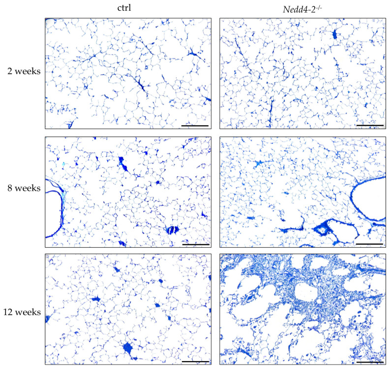
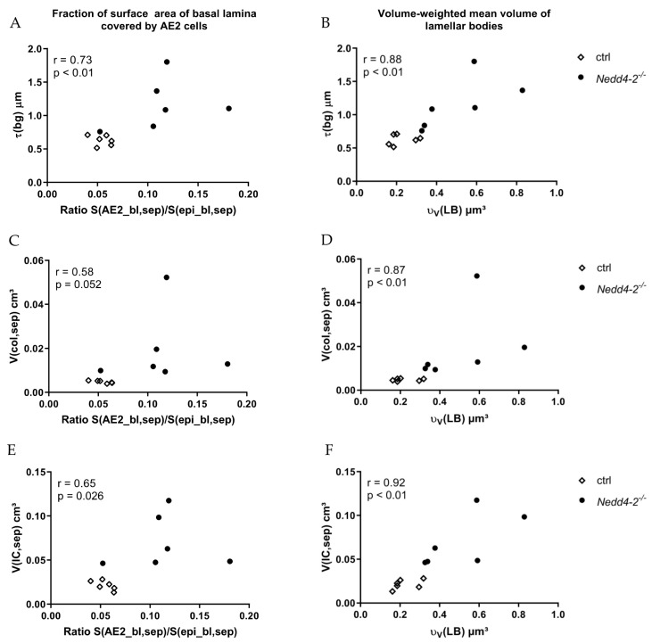
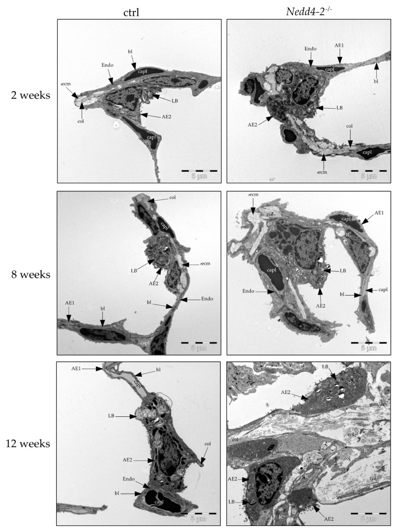
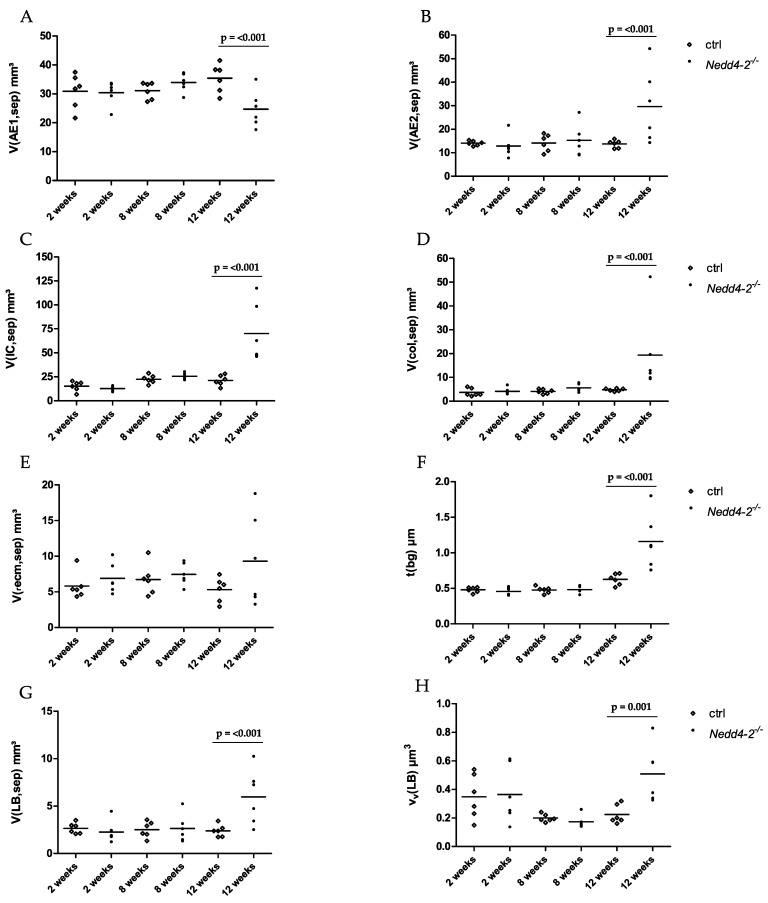
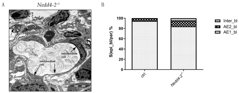
Our previous study showed that in adult mice, conditional -deficiency in club and alveolar epithelial type II (AE2) cells results in impaired mucociliary clearance, accumulation of and progressive, terminal pulmonary fibrosis within 16 weeks. In the present study, we investigated ultrastructural alterations of the alveolar epithelium in relation to interstitial remodeling in alveolar septa as a function of disease progression. Two, eight and twelve weeks after induction of knockout, lungs were fixed and subjected to design-based stereological investigation at the light and electron microscopic level. Quantitative data did not show any abnormalities until 8 weeks compared to controls. At 12 weeks, however, volume of septal wall tissue increased while volume of acinar airspace and alveolar surface area significantly decreased. Volume and surface area of alveolar epithelial type I cells were reduced, which could not be compensated by a corresponding increase of AE2 cells. The volume of collagen fibrils in septal walls increased and was linked with an increase in blood-gas barrier thickness. A high correlation between parameters reflecting interstitial remodeling and abnormal AE2 cell ultrastructure could be established. Taken together, abnormal regeneration of the alveolar epithelium is correlated with interstitial septal wall remodeling.
我们之前的研究表明,在成年小鼠中,条件性敲除 club 和肺泡上皮细胞 II 型(AE2)会导致黏液纤毛清除功能受损、积聚和进行性终末性肺纤维化,这一过程在 16 周内发生。在本研究中,我们研究了肺泡上皮的超微结构改变与肺泡隔间质重塑之间的关系,以探讨其作为疾病进展的功能。在诱导 基因敲除后 2、8 和 12 周,固定肺组织并在光镜和电镜下进行基于设计的体视学研究。与对照组相比,直到 8 周时定量数据才显示出任何异常。然而,在 12 周时,隔壁组织体积增加,而腺泡腔和肺泡表面积显著减少。肺泡上皮细胞 I 型的体积和表面积减少,这不能通过相应增加 AE2 细胞来补偿。隔壁胶原纤维的体积增加,与气血屏障厚度增加有关。可以建立反映间质重塑和异常 AE2 细胞超微结构之间的高相关性参数。总之,肺泡上皮的异常再生与间质隔壁重塑有关。