Department of Psychiatry, Icahn School of Medicine at Mount Sinai, New York, New York, USA.
Seaver Autism Center for Research and Treatment, Icahn School of Medicine at Mount Sinai, New York, New York, USA.
Autism Res. 2021 Sep;14(9):1837-1842. doi: 10.1002/aur.2568. Epub 2021 Jul 27.
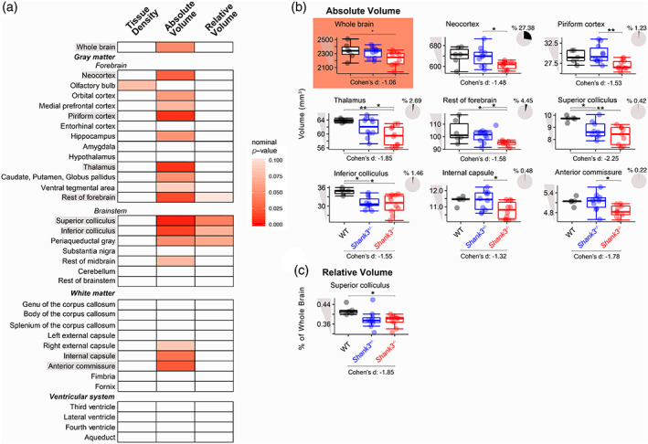
Mutations and deletions in the SHANK3 gene cause the major neurodevelopmental features of Phelan-McDermid syndrome (PMS), which is characterized by intellectual disability, autism spectrum disorder, and sensory hyporeactivity. SHANK3 encodes a key structural component of excitatory synapses important for synaptogenesis. Clinical assessments and limited brain imaging studies of patients with PMS have uncovered regional volume reductions and white matter thinning. While these impairments have been replicated ex vivo in pups of a rat model, brain structure has not been assessed in rats in vivo or in adults. We assessed the brain structure of heterozygous and homozygous adult Shank3-deficient male rats in comparison to wild-type littermates with magnetic resonance imaging using both anatomical assessments and diffusion tensor imaging (DTI). Shank3-deficient rats showed a reduction in overall brain size and the absolute volume of the neocortex, piriform cortex, thalamus, forebrain, inferior and superior colliculi, internal capsule, and anterior commissure. The superior colliculus was decreased in relative volume. DTI revealed that axial diffusion and fractional anisotropy were reduced in the external capsule and mean diffusion was increased in the fornix, suggesting that restriction of diffusion perpendicular to the axis of the axonal fibers was impaired in these white matter tracts. Therefore, Shank3-deficient rats replicate the reduced brain volume and altered white matter phenotypes present in PMS. Our results indicate that the loss of a glutamatergic synaptic protein, Shank3, has structural consequences at the level of the whole brain. The brain regions that were altered represent potential cross-species structural biomarkers that warrant further study.
SHANK3 基因的突变和缺失导致了 Phelan-McDermid 综合征(PMS)的主要神经发育特征,其特征为智力障碍、自闭症谱系障碍和感觉反应迟钝。SHANK3 编码兴奋性突触的关键结构组成部分,对于突触发生很重要。对 PMS 患者的临床评估和有限的脑成像研究揭示了区域性体积减少和白质变薄。虽然这些损伤在 PMS 的大鼠模型的幼崽中得到了复制,但在体内的大鼠或成年大鼠中尚未评估脑结构。我们使用磁共振成像对杂合子和纯合子成年 Shank3 缺陷型雄性大鼠与野生型同窝仔鼠进行了脑结构评估,包括解剖学评估和弥散张量成像(DTI)。Shank3 缺陷型大鼠的总脑体积和新皮层、梨状皮层、丘脑、前脑、上丘和下丘、内囊和前连合的绝对体积减小。上丘的相对体积减小。DTI 显示外囊的轴向扩散和各向异性分数降低,穹窿的平均扩散增加,表明这些白质束中与轴突纤维垂直的扩散受限受损。因此,Shank3 缺陷型大鼠复制了 PMS 中存在的脑体积减小和白质表型改变。我们的结果表明,谷氨酸能突触蛋白 Shank3 的缺失会对整个大脑的结构产生影响。改变的脑区代表了潜在的跨物种结构生物标志物,值得进一步研究。