Chen Jun, Patel Toral R, Pinho Marco C, Choi Changho, Harrison Crystal E, Baxter Jeannie D, Derner Kelley, Pena Salvador, Liticker Jeff, Raza Jaffar, Hall Ronald G, Reed Galen D, Cai Chunyu, Hatanpaa Kimmo J, Bankson James A, Bachoo Robert M, Malloy Craig R, Mickey Bruce E, Park Jae Mo
Advanced Imaging Research Center, The University of Texas Southwestern Medical Center, Dallas, Texas, USA.
Department of Neurosurgery, The University of Texas Southwestern Medical Center, Dallas, Texas, USA.
Neurooncol Adv. 2021 Jun 28;3(1):vdab092. doi: 10.1093/noajnl/vdab092. eCollection 2021 Jan-Dec.
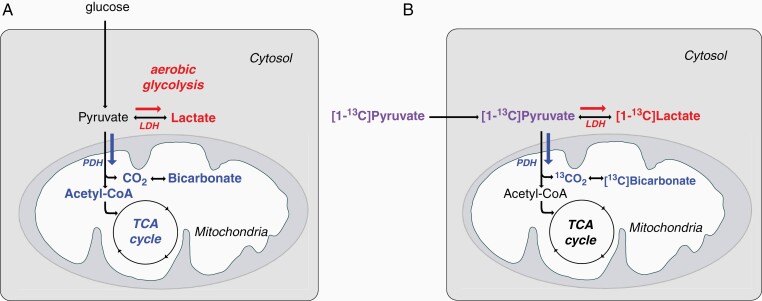
Glioblastoma remains incurable despite treatment with surgery, radiation therapy, and cytotoxic chemotherapy, prompting the search for a metabolic pathway unique to glioblastoma cells.C MR spectroscopic imaging with hyperpolarized pyruvate can demonstrate alterations in pyruvate metabolism in these tumors.
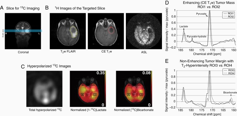
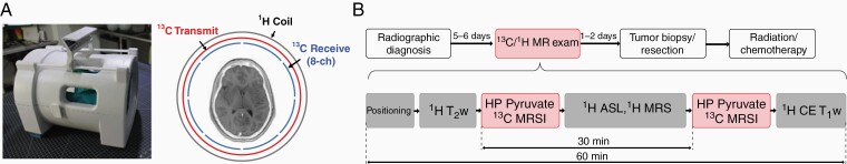
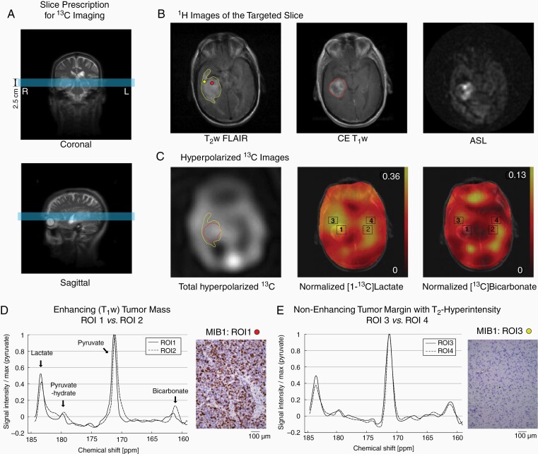
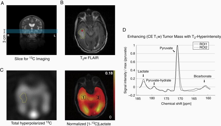
Three patients with diagnostic MRI suggestive of a glioblastoma were scanned at 3 T 1-2 days prior to tumor resection using a C/H dual-frequency RF coil and a C/H-integrated MR protocol, which consists of a series of H MR sequences (T FLAIR, arterial spin labeling and contrast-enhanced [CE] T) and C spectroscopic imaging with hyperpolarized [1-C]pyruvate. Dynamic spiral chemical shift imaging was used for C data acquisition. Surgical navigation was used to correlate the locations of tissue samples submitted for histology with the changes seen on the diagnostic MR scans and the C spectroscopic images.
Each tumor was histologically confirmed to be a WHO grade IV glioblastoma with isocitrate dehydrogenase wild type. Total hyperpolarized C signals detected near the tumor mass reflected altered tissue perfusion near the tumor. For each tumor, a hyperintense [1-C]lactate signal was detected both within CE and T-FLAIR regions on the H diagnostic images ( = .008). [C]bicarbonate signal was maintained or decreased in the lesion but the observation was not significant ( = .3).
Prior to surgical resection, C MR spectroscopic imaging with hyperpolarized pyruvate reveals increased lactate production in regions of histologically confirmed glioblastoma.
尽管胶质母细胞瘤接受了手术、放射治疗和细胞毒性化疗,但仍无法治愈,这促使人们寻找胶质母细胞瘤细胞特有的代谢途径。使用超极化丙酮酸的碳磁共振波谱成像可以显示这些肿瘤中丙酮酸代谢的改变。
3例诊断性MRI提示为胶质母细胞瘤的患者在肿瘤切除术前1 - 2天,使用碳/氢双频射频线圈和碳/氢一体化MR协议在3T下进行扫描,该协议包括一系列氢MR序列(液体衰减反转恢复序列、动脉自旋标记和对比增强[CE] T1加权像)以及超极化[1-13C]丙酮酸的碳波谱成像。动态螺旋化学位移成像用于碳数据采集。手术导航用于将送检组织学检查的组织样本位置与诊断性MR扫描和碳波谱图像上看到的变化相关联。
每个肿瘤经组织学证实为异柠檬酸脱氢酶野生型的世界卫生组织IV级胶质母细胞瘤。在肿瘤块附近检测到的总超极化碳信号反映了肿瘤附近组织灌注的改变。对于每个肿瘤,在氢诊断图像上的CE和液体衰减反转恢复区域内均检测到高强度的[1-13C]乳酸信号(P = 0.008)。病变内的[13C]碳酸氢盐信号保持或降低,但观察结果不显著(P = 0.3)。
在手术切除前,使用超极化丙酮酸的碳磁共振波谱成像显示,在组织学证实的胶质母细胞瘤区域乳酸生成增加。