Kim Hyun Sik, Park Young Han, Lee Heui Seung, Kwon Mi Jung, Song Joon Ho, Chang In Bok
Department of Neurosurgery, Hallym University Sacred Heart Hospital, Anyang, Korea.
Department of Obstetrics and Gynecology, Hallym University Sacred Heart Hospital, Anyang, Korea.
J Korean Neurosurg Soc. 2021 Sep;64(5):716-725. doi: 10.3340/jkns.2021.0068. Epub 2021 Aug 11.
The anti-tumor effect of the beta-adrenergic receptor antagonist propranolol in breast cancer is well known; however, its activity in glioblastoma is not well-evaluated. The Notch-Hes pathway is known to regulate cell differentiation, proliferation, and apoptosis. We investigated the effect of propranolol to human glioblastoma cell lines, and the role of Notch and Hes signaling in this process.
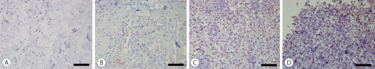
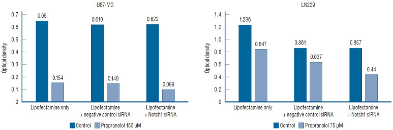
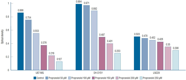
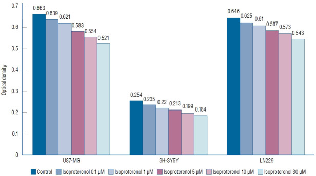
We performed immunohistochemical staining on 31 surgically resected primary human glioblastoma tissues. We also used glioblastoma cell lines of U87-MG, LN229, and neuroblastoma cell line of SH-SY5Y in this study. The effect of propranolol and isoproterenol on cell proliferation was evaluated using the MTT assay (absorbance 570 nm). The impact of propranolol on gene expression (Notch and Hes) was evaluated using real-time polymerase chain reaction (RT-PCR, whereas protein levels of Notch1 and Hes1 were measured using Western blotting (WB), simultaneously. Small interfering RNA (siRNA) was used to suppress the Notch gene to investigate its role in the proliferation of glioblastoma.
Propranolol and isoproterenol caused a dose-dependent decrease in cell proliferation (MTT assay). RT-PCR showed an increase in Notch1 and Hes1 expression by propranolol, whereas WB demonstrated increase in Notch1 protein, but a decrease in Hes1 by propranolol. The proliferation of U87-MG and LN229 was not significantly suppressed after transfection with Notch siRNA.
These results demonstrated that propranolol suppressed the proliferation of glioblastoma cell lines and neuroblastoma cell line, and Hes1 was more closely involved than Notch1 was in glioblastoma proliferation.
β-肾上腺素能受体拮抗剂普萘洛尔在乳腺癌中的抗肿瘤作用已广为人知;然而,其在胶质母细胞瘤中的活性尚未得到充分评估。已知Notch-Hes信号通路可调节细胞分化、增殖和凋亡。我们研究了普萘洛尔对人胶质母细胞瘤细胞系的作用,以及Notch和Hes信号在这一过程中的作用。
我们对31例手术切除的原发性人胶质母细胞瘤组织进行了免疫组化染色。在本研究中,我们还使用了U87-MG、LN229胶质母细胞瘤细胞系以及SH-SY5Y神经母细胞瘤细胞系。使用MTT法(吸光度570nm)评估普萘洛尔和异丙肾上腺素对细胞增殖的影响。使用实时聚合酶链反应(RT-PCR)评估普萘洛尔对基因表达(Notch和Hes)的影响,同时使用蛋白质印迹法(WB)检测Notch1和Hes1的蛋白水平。使用小干扰RNA(siRNA)抑制Notch基因,以研究其在胶质母细胞瘤增殖中的作用。
普萘洛尔和异丙肾上腺素导致细胞增殖呈剂量依赖性下降(MTT法)。RT-PCR显示普萘洛尔使Notch1和Hes1表达增加,而WB显示普萘洛尔使Notch1蛋白增加,但使Hes1蛋白减少。用Notch siRNA转染后,U87-MG和LN229的增殖未受到显著抑制。
这些结果表明,普萘洛尔抑制了胶质母细胞瘤细胞系和神经母细胞瘤细胞系的增殖,并且在胶质母细胞瘤增殖中,Hes1比Notch1参与程度更高。