Hu Lingfei, Liu Yinping, Wei Chaobing, Jin Huixiang, Mei Lixin, Wu Changfan
Department of Ophthalmology, Yijishan Hospital of Wannan Medical College, Wuhu, Anhui Province, People's Republic of China.
Diabetes Metab Syndr Obes. 2021 Aug 4;14:3471-3483. doi: 10.2147/DMSO.S307771. eCollection 2021.
In the present study, we performed bioinformatics studies and in vitro functional assays to explore the underlying role of serpin family H member 1 (SERPINH1) in the diabetic retinopathy.
Common differentially expressed genes (DEGs) between diabetic retinal tissues and normal retinal tissues were analyzed using Gene Expression Omnibus (GEO) database. The proliferation and migration of human retinal endothelial cells (HRECs) was evaluated by MTS, EdU and wound healing assays, respectively; the miRNA and mRNAs expression levels of hub genes in HRECs were determined using quantitative real-time PCR (qRT-PCR). Protein levels were determined using a Western blot assay.
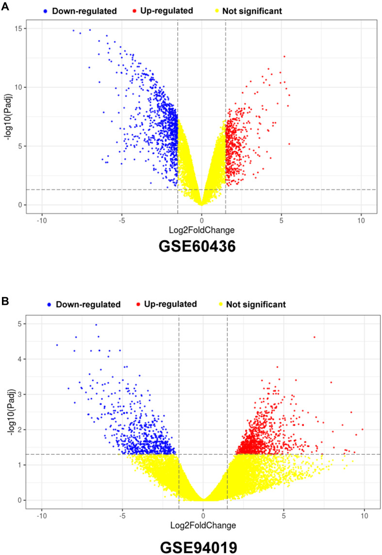
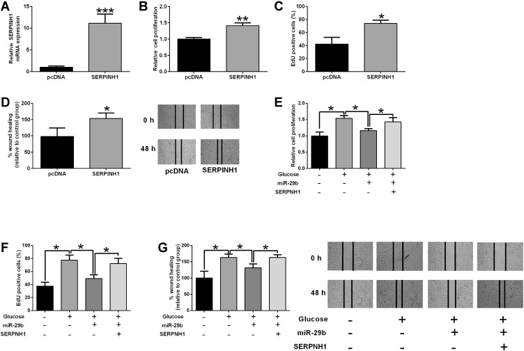
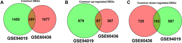
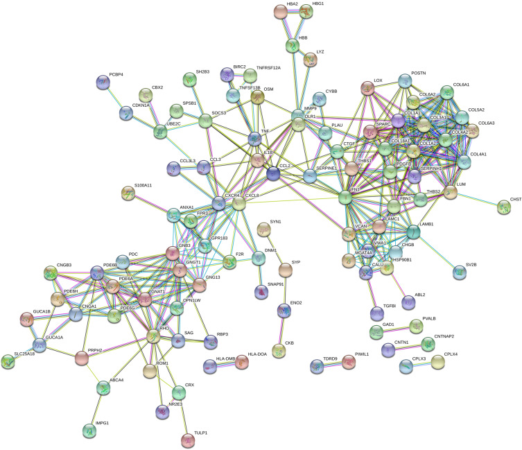
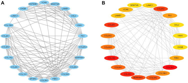
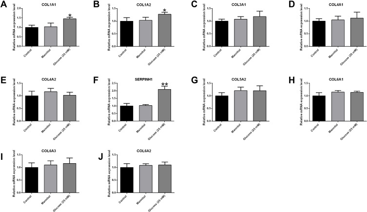
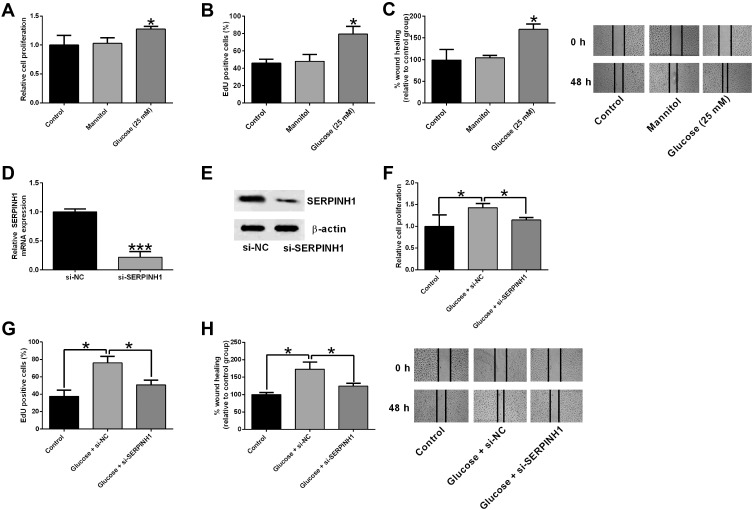
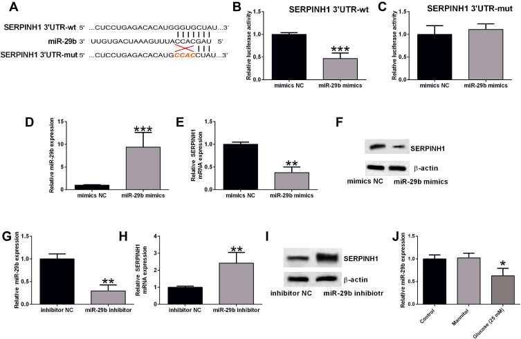
A total of 189 common DEGs were screened between two GEO datasets (GSE60436 and GSE94019), and ten potential hub genes that may link to the progression of diabetic retinopathy were detected. The qRT-PCR results showed that collagen, type I, alpha 1 (COL1A1), Collagen, type I, alpha 2 (COL1A2) and serpin family H member 1 (SERPINH1) mRNA expression levels were up-regulated in the HRECs after being exposed to high glucose for 48 h. Silence of SERPINH1 repressed the high glucose-induced increase in proliferation and migration of HRECs. SERPINH1 was a target of miR-29b and was suppressed by miR-29 in HRECs. SERPINH1 overexpression promoted HREC proliferation and migration. Furthermore, miR-29b suppressed HREC proliferation and migration under high-glucose stimulation, which was significantly attenuated by enforced expression of SERPINH1.
In conclusion, by performing the integrated bioinformatics analysis, the present study suggested that 3 hub genes (COL1A1, COL1A2 and SERPINH1) may be associated with diabetic retinopathy pathophysiology. Further mechanistic studies indicated that miR-29b/SERPINH1 signaling participated in high glucose-induced enhancement in the proliferation and migration of HRECs.
在本研究中,我们进行了生物信息学研究和体外功能试验,以探究丝氨酸蛋白酶抑制剂家族H成员1(SERPINH1)在糖尿病视网膜病变中的潜在作用。
使用基因表达综合数据库(GEO)分析糖尿病视网膜组织和正常视网膜组织之间的常见差异表达基因(DEG)。分别通过MTS、EdU和伤口愈合试验评估人视网膜内皮细胞(HREC)的增殖和迁移;使用定量实时PCR(qRT-PCR)测定HREC中枢纽基因的miRNA和mRNA表达水平。使用蛋白质印迹法测定蛋白质水平。
在两个GEO数据集(GSE60436和GSE94019)之间共筛选出189个常见DEG,并检测到10个可能与糖尿病视网膜病变进展相关的潜在枢纽基因。qRT-PCR结果显示,在暴露于高糖48小时后,HREC中I型胶原α1链(COL1A1)、I型胶原α2链(COL1A2)和丝氨酸蛋白酶抑制剂家族H成员1(SERPINH1)的mRNA表达水平上调。沉默SERPINH1可抑制高糖诱导的HREC增殖和迁移增加。SERPINH1是miR-29b的靶标,在HREC中被miR-29抑制。SERPINH1过表达促进HREC增殖和迁移。此外,miR-29b在高糖刺激下抑制HREC增殖和迁移,而SERPINH1的强制表达可显著减弱这种抑制作用。
总之,通过进行综合生物信息学分析,本研究表明3个枢纽基因(COL1A1、COL1A2和SERPINH1)可能与糖尿病视网膜病变的病理生理学相关。进一步的机制研究表明,miR-29b/SERPINH1信号通路参与了高糖诱导的HREC增殖和迁移增强。