Institut Pasteur de Montevideo, 2020, Montevideo, Uruguay.
Department of Neurology, University of Alabama, Birmingham, AL, 35294, USA.
Acta Neuropathol Commun. 2021 Aug 13;9(1):136. doi: 10.1186/s40478-021-01241-3.
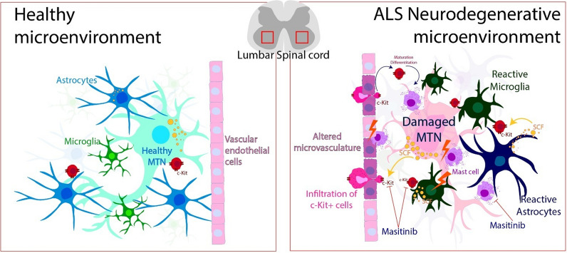
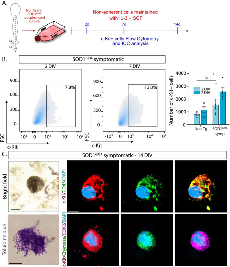
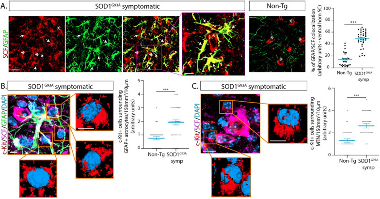
Degeneration of motor neurons, glial cell reactivity, and vascular alterations in the CNS are important neuropathological features of amyotrophic lateral sclerosis (ALS). Immune cells trafficking from the blood also infiltrate the affected CNS parenchyma and contribute to neuroinflammation. Mast cells (MCs) are hematopoietic-derived immune cells whose precursors differentiate upon migration into tissues. Upon activation, MCs undergo degranulation with the ability to increase vascular permeability, orchestrate neuroinflammation and modulate the neuroimmune response. However, the prevalence, pathological significance, and pharmacology of MCs in the CNS of ALS patients remain largely unknown. In autopsy ALS spinal cords, we identified for the first time that MCs express c-Kit together with chymase, tryptase, and Cox-2 and display granular or degranulating morphology, as compared with scarce MCs in control cords. In ALS, MCs were mainly found in the niche between spinal motor neuron somas and nearby microvascular elements, and they displayed remarkable pathological abnormalities. Similarly, MCs accumulated in the motor neuron-vascular niche of ALS murine models, in the vicinity of astrocytes and motor neurons expressing the c-Kit ligand stem cell factor (SCF), suggesting an SCF/c-Kit-dependent mechanism of MC differentiation from precursors. Mechanistically, we provide evidence that fully differentiated MCs in cell cultures can be generated from the murine ALS spinal cord tissue, further supporting the presence of c-Kit+ MC precursors. Moreover, intravenous administration of bone marrow-derived c-Kit+ MC precursors infiltrated the spinal cord in ALS mice but not in controls, consistent with aberrant trafficking through a defective microvasculature. Pharmacological inhibition of c-Kit with masitinib in ALS mice reduced the MC number and the influx of MC precursors from the periphery. Our results suggest a previously unknown pathogenic mechanism triggered by MCs in the ALS motor neuron-vascular niche that might be targeted pharmacologically.
运动神经元变性、神经胶质细胞反应和中枢神经系统的血管改变是肌萎缩侧索硬化症(ALS)的重要神经病理学特征。从血液中迁移而来的免疫细胞也会渗透到受影响的中枢神经系统实质中,并有助于神经炎症。肥大细胞(MCs)是造血衍生的免疫细胞,其前体在迁移到组织中时会分化。一旦被激活,MC 就会脱颗粒,从而增加血管通透性、协调神经炎症并调节神经免疫反应。然而,MC 在 ALS 患者中枢神经系统中的流行程度、病理意义和药理学仍然知之甚少。在 ALS 患者的尸检脊髓中,我们首次发现 MC 表达 c-Kit 以及糜蛋白酶、类胰蛋白酶和 Cox-2,并表现出颗粒状或脱颗粒状形态,而对照脊髓中的 MC 则很少。在 ALS 中,MC 主要存在于脊髓运动神经元体和附近微血管之间的间隙中,并表现出明显的病理异常。同样,MC 在 ALS 动物模型的运动神经元-血管间隙中积累,靠近表达 c-Kit 配体干细胞因子(SCF)的星形胶质细胞和运动神经元,表明存在从前体分化为 MC 的 SCF/c-Kit 依赖性机制。从机制上讲,我们提供的证据表明,可以从鼠 ALS 脊髓组织中培养出完全分化的 MC,进一步支持存在 c-Kit+MC 前体。此外,静脉注射骨髓衍生的 c-Kit+MC 前体可渗透到 ALS 小鼠的脊髓中,但在对照组中不会,这与通过有缺陷的微血管异常渗透有关。在 ALS 小鼠中用 masitinib 抑制 c-Kit 可减少 MC 数量和来自外周的 MC 前体的涌入。我们的研究结果表明,在 ALS 运动神经元-血管间隙中,MC 触发了一个以前未知的致病机制,该机制可能在药理学上被靶向。