Minoda Alexandre Makoto, Ferreira Fernando Dos Santos, Santos Karllos Diego Ribeiro, Leão Cristiano de Souza, Silva Eduardo Just da Costa E, de Melo-Leite Andréa Farias
Instituto de Medicina Integral Professor Fernando Figueira (IMIP), Recife, PE, Brasil.
Universidade Federal de Pernambuco (UFPE), Recife, PE, Brazil.
Radiol Bras. 2021 Jul-Aug;54(4):270-276. doi: 10.1590/0100-3984.2020.0108.
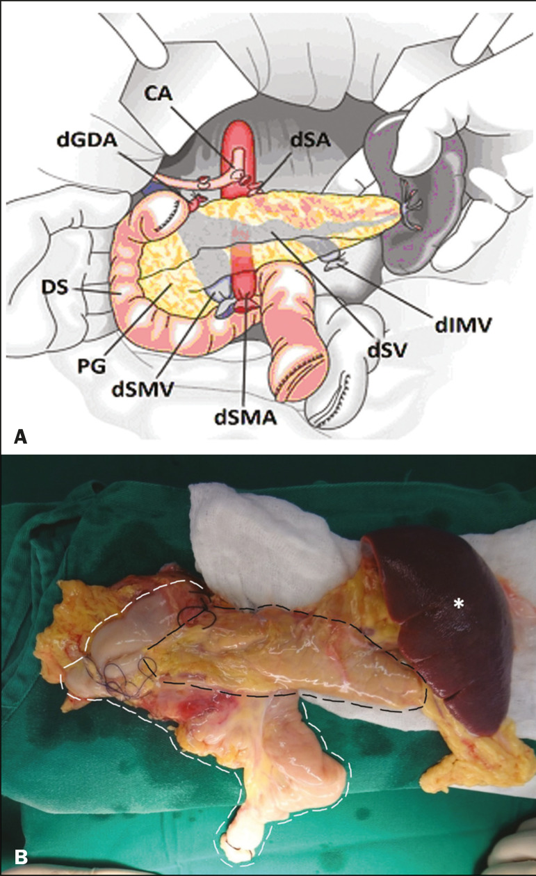
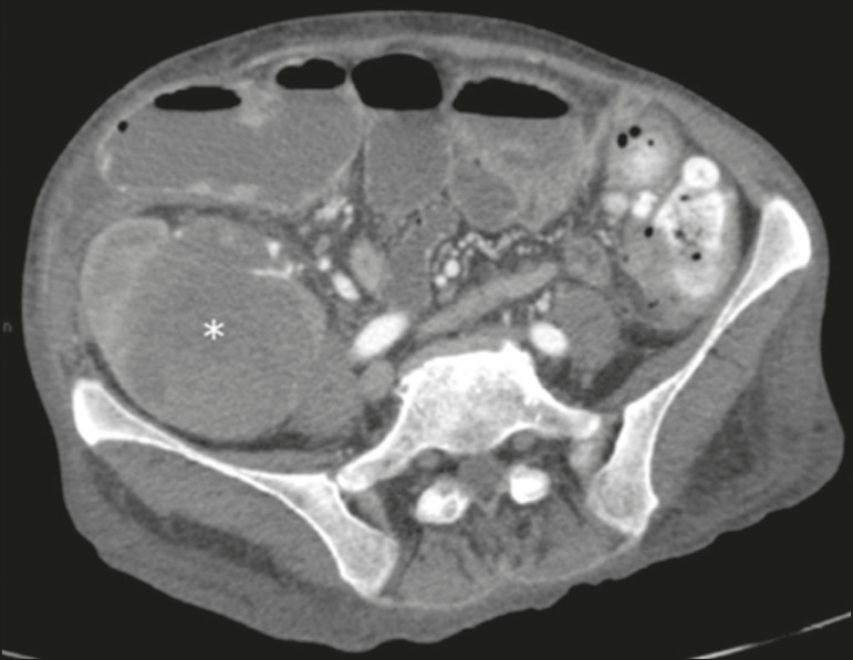
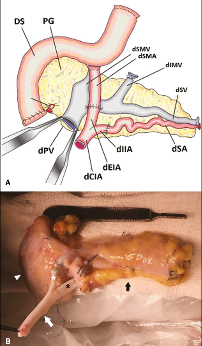
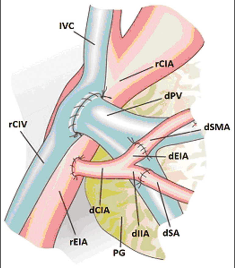
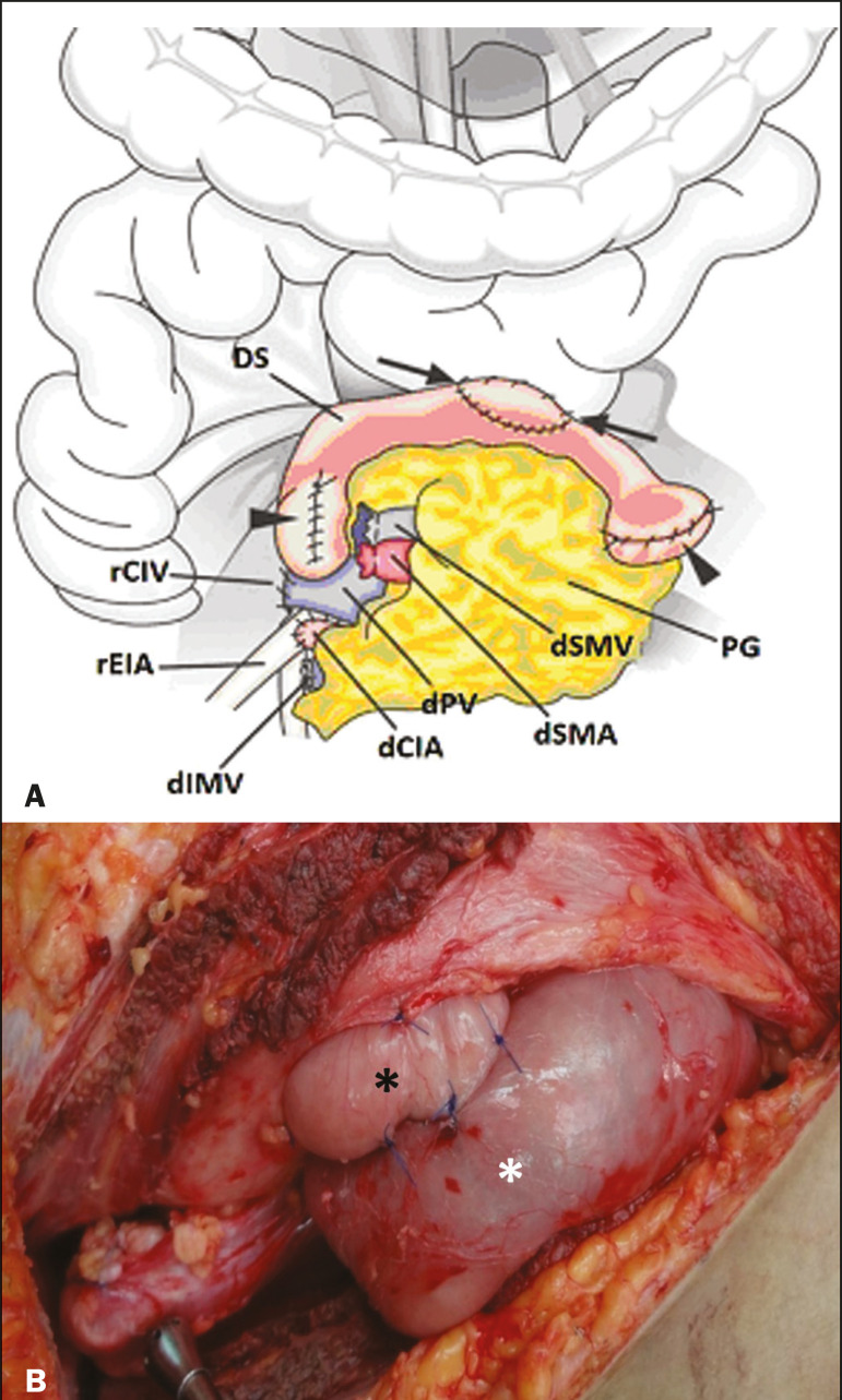
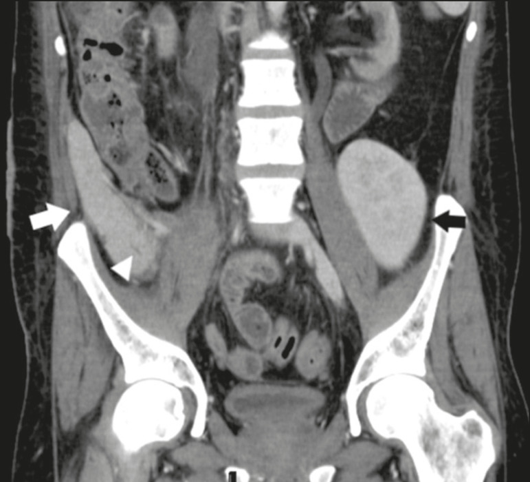
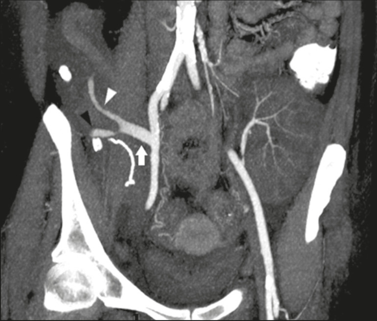
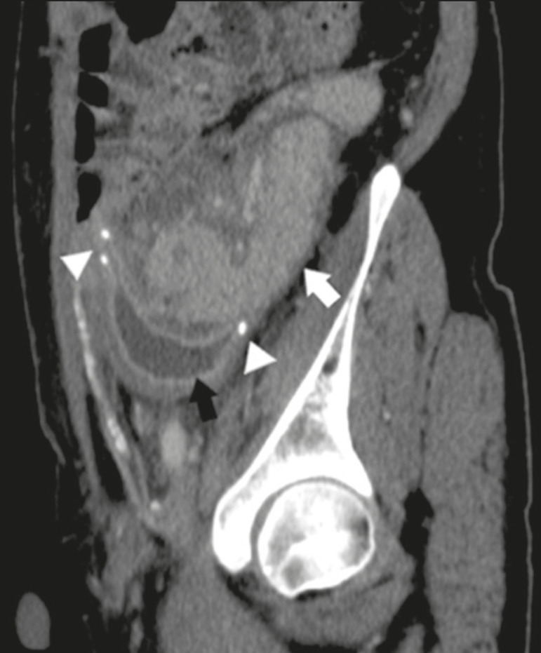
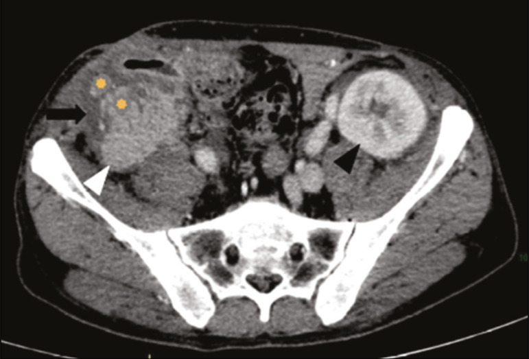
Pancreas transplantation is a well-established treatment for patients with complicated diabetes mellitus and advanced renal failure. The most common procedure is simultaneous pancreas-kidney transplantation, in which the pancreas graft is positioned in the right pelvic region and the kidney graft is positioned in the left iliac fossa. Various imaging methods are used for the post-transplantation evaluation of the graft parenchyma and vascular anatomy, as well as for the identification of possible complications. As the number of cases increases, it is fundamental that radiologists understand the surgical procedure and the postoperative anatomy, as well as to recognize the possible postoperative complications and their imaging aspects, with the aim of providing the best guidance in the postoperative management of transplant recipients.
胰腺移植是治疗复杂糖尿病和晚期肾衰竭患者的一种成熟疗法。最常见的手术是胰肾联合移植,其中胰腺移植物置于右盆腔区域,肾移植物置于左髂窝。各种成像方法用于移植后对移植物实质和血管解剖结构的评估,以及用于识别可能的并发症。随着病例数量的增加,放射科医生了解手术过程和术后解剖结构,以及识别可能的术后并发症及其影像学表现至关重要,目的是为移植受者的术后管理提供最佳指导。