Department of Radiation Physics and Technology, Southern TOHOKU Proton Therapy Center, Koriyama, Japan.
School of Health Sciences, Fukushima Medical University, Fukushima, Japan.
J Appl Clin Med Phys. 2021 Sep;22(9):298-306. doi: 10.1002/acm2.13391. Epub 2021 Aug 17.
Anatomical changes, such as shrinkage and aeration, can affect dose distribution in proton therapy (PT) for maxillary sinus carcinoma (MSC). These changes can affect the dose to the target and organs at risk (OARs); however, when these changes occur during PT is unclear. This study aimed to investigate the dosimetric impact of anatomical changes during PT.
Fifteen patients with MSC were enrolled in this study. Initial PT plans were generated based on initial computed tomography (CT) images. Several repeat CT images were obtained to confirm anatomical changes during PT. Evaluation PT plans were generated by copying initial PT plans to repeat CT images. The dose differences of the target and OARs were evaluated by comparing both the plans.
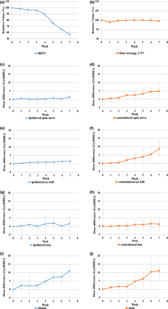
At 3-4 weeks after the initiation of PT, the target volume reduced by approximately 10% as compared with the initial volume. Consequently, the target volumes gradually varied until the end of treatment. The value of V (volume that received 95% of the prescription dose) in the clinical target volume of the evaluation PT plan was similar to that of the initial PT plan. However, the dose to OARs, such as the contralateral optic nerve, contralateral eyeball, brainstem, and optic chiasm, increased significantly from the middle to the later phases of the treatment course. In contrast, there was a slight dose difference in the ipsilateral optic apparatus.
The trend analysis in this study showed that anatomical changes appeared 3-4 weeks after the start of PT, and the dose to the OARs tended to increase. Therefore, it is recommended to check the status of tumor 3-4 weeks after the start of treatment to avoid the deterioration of dose distribution due to these changes.
解剖结构的变化,如收缩和充气,会影响上颌窦癌(MSC)质子治疗(PT)中的剂量分布。这些变化会影响靶区和危及器官(OAR)的剂量;然而,这些变化在 PT 期间何时发生尚不清楚。本研究旨在探讨 PT 期间解剖结构变化对剂量学的影响。
本研究纳入了 15 例 MSC 患者。初始 PT 计划基于初始计算机断层扫描(CT)图像生成。获得了几次重复 CT 图像以确认 PT 期间的解剖变化。通过将初始 PT 计划复制到重复 CT 图像上,生成评估 PT 计划。通过比较这两种计划来评估靶区和 OAR 的剂量差异。
PT 开始后 3-4 周,与初始体积相比,靶区体积减少了约 10%。因此,靶区体积逐渐变化,直到治疗结束。评估 PT 计划的靶区临床靶区体积(V)值与初始 PT 计划相似。然而,从治疗过程的中期到后期,OAR 的剂量,如对侧视神经、对侧眼球、脑干和视交叉,显著增加。相比之下,同侧视神经器官的剂量差异较小。
本研究的趋势分析表明,解剖结构的变化在 PT 开始后 3-4 周出现,OAR 的剂量趋于增加。因此,建议在治疗开始后 3-4 周检查肿瘤的状况,以避免由于这些变化导致剂量分布恶化。