Rolfes Leoni, Schulte-Mecklenbeck Andreas, Schreiber Stefanie, Vielhaber Stefan, Herty Michael, Marten Anika, Pfeuffer Steffen, Ruck Tobias, Wiendl Heinz, Gross Catharina C, Meuth Sven G, Boentert Matthias, Pawlitzki Marc
Department of Neurology with Institute of Translational Neurology, University Hospital Münster, Münster 48149, Germany.
Department of Neurology, University Hospital Düsseldorf, Heinrich-Heine-University, Düsseldorf 40225, Germany.
Brain Commun. 2021 Jul 14;3(3):fcab157. doi: 10.1093/braincomms/fcab157. eCollection 2021.
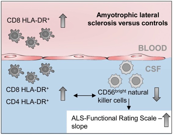
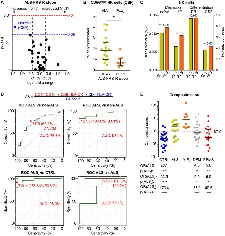
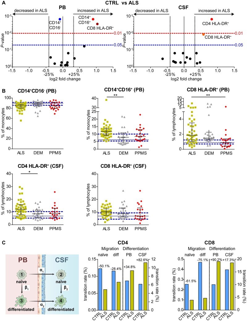
Several studies suggest a role for the peripheral immune system in the pathophysiology of amyotrophic lateral sclerosis. However, comprehensive studies investigating the intrathecal immune system in amyotrophic lateral sclerosis are rare. To elucidate whether compartment-specific inflammation contributes to amyotrophic lateral sclerosis pathophysiology, we here investigated intrathecal and peripheral immune profiles in amyotrophic lateral sclerosis patients and compared them with controls free of neurological disorders (controls) and patients with dementia or primary progressive multiple sclerosis. Routine CSF parameters were examined in 308 patients, including 132 amyotrophic lateral sclerosis patients. In a subgroup of 41 amyotrophic lateral sclerosis patients, extensive flow-cytometric immune cell profiling in peripheral blood and CSF was performed and compared with data from 26 controls, 25 dementia and 21 multiple sclerosis patients. Amyotrophic lateral sclerosis patients presented with significantly altered proportions of monocyte subsets in peripheral blood and increased frequencies of CD4 and CD8 T cells expressing the activation marker HLA-DR in peripheral blood (CD8) and CSF (CD4 and CD8) compared with controls. While dementia and multiple sclerosis patients exhibited a comparable increase in intrathecal CD8 T-cell activation, CD8 T-cell activation in the peripheral blood in amyotrophic lateral sclerosis was higher than in multiple sclerosis patients. Furthermore, intrathecal CD4 T-cell activation in amyotrophic lateral sclerosis surpassed levels in dementia patients. Intrathecal T-cell activation resulted from activation rather than transmigration of activated T cells from the blood. While T-cell activation did not correlate with amyotrophic lateral sclerosis progression, patients with rapid disease progression showed reduced intrathecal levels of immune-regulatory CD56 natural killer cells. The integration of these parameters into a composite score facilitated the differentiation of amyotrophic lateral sclerosis patients from patients of all other cohorts. To conclude, alterations in peripheral monocyte subsets, as well as increased peripheral and intrathecal activation of CD4 and CD8 T cells concomitant with diminished immune regulation by CD56 natural killer cells, suggest an involvement of these immune cells in amyotrophic lateral sclerosis pathophysiology.
多项研究表明外周免疫系统在肌萎缩侧索硬化症的病理生理学中发挥作用。然而,针对肌萎缩侧索硬化症鞘内免疫系统的全面研究却很罕见。为了阐明特定腔室的炎症是否导致肌萎缩侧索硬化症的病理生理学,我们在此研究了肌萎缩侧索硬化症患者的鞘内和外周免疫特征,并将其与无神经系统疾病的对照组(对照)以及患有痴呆症或原发性进行性多发性硬化症的患者进行比较。对308名患者进行了常规脑脊液参数检查,其中包括132名肌萎缩侧索硬化症患者。在41名肌萎缩侧索硬化症患者的亚组中,对外周血和脑脊液进行了广泛的流式细胞术免疫细胞分析,并与26名对照、25名痴呆症患者和21名多发性硬化症患者的数据进行了比较。与对照组相比,肌萎缩侧索硬化症患者外周血中单核细胞亚群比例显著改变,外周血(CD8)和脑脊液(CD4和CD8)中表达活化标志物HLA-DR的CD4和CD8 T细胞频率增加。虽然痴呆症和多发性硬化症患者鞘内CD8 T细胞活化程度相当,但肌萎缩侧索硬化症患者外周血中的CD8 T细胞活化高于多发性硬化症患者。此外,肌萎缩侧索硬化症患者鞘内CD4 T细胞活化超过了痴呆症患者的水平。鞘内T细胞活化是由活化T细胞的活化而非从血液中迁移所致。虽然T细胞活化与肌萎缩侧索硬化症进展无关,但疾病进展迅速的患者鞘内免疫调节性CD56自然杀伤细胞水平降低。将这些参数整合为一个综合评分有助于区分肌萎缩侧索硬化症患者与所有其他队列的患者。总之,外周单核细胞亚群的改变,以及CD4和CD8 T细胞外周和鞘内活化增加,同时CD56自然杀伤细胞的免疫调节作用减弱,表明这些免疫细胞参与了肌萎缩侧索硬化症的病理生理学过程。