Mathias Maxwell, Taylor Joann, Mendralla Elizabeth, Perez Marta
Division of Neonatology, Department of Pediatrics, Northwestern University Feinberg School of Medicine, Chicago, IL 60611, USA.
Ann & Robert H. Lurie Children's Hospital of Chicago, Chicago, IL 60611, USA.
Antioxidants (Basel). 2021 Aug 1;10(8):1236. doi: 10.3390/antiox10081236.
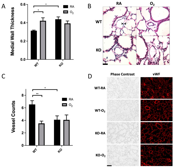
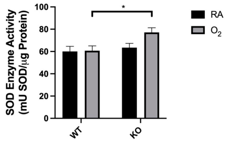
Bronchopulmonary dysplasia (BPD) is a common lung disease affecting premature infants that develops after exposure to supplemental oxygen and reactive oxygen intermediates. Extracellular superoxide dismutase (SOD3) is an enzyme that processes superoxide radicals and has been shown to facilitate vascular endothelial growth factor (VEGF) and nitric oxide (NO) signaling in vascular endothelium. We utilized a mouse model of neonatal hyperoxic lung injury and SOD3 knockout (KO) mice to evaluate its function during chronic hyperoxia exposure. Wild-type age-matched neonatal C57Bl/6 (WT) and SOD3 (KO) mice were placed in normoxia (21% FiO, RA) or chronic hyperoxia (75% FiO, O) within 24 h of birth for 14 days continuously and then euthanized. Lungs were harvested for histologic evaluation, as well as comparison of antioxidant enzyme expression, SOD activity, VEGF expression, and portions of the NO signaling pathway. Surprisingly, KO-O mice survived without additional alveolar simplification, microvascular remodeling, or nuclear oxidation when compared to WT-O mice. KO-O mice had increased total SOD activity and increased VEGF expression when compared to WT-O mice. No genotype differences were noted in intracellular antioxidant enzyme expression or the NO signaling pathway. These results demonstrate that SOD3 KO mice can survive prolonged hyperoxia without exacerbation of alveolar or vascular phenotype.
支气管肺发育不良(BPD)是一种常见的肺部疾病,影响早产儿,在暴露于补充氧气和活性氧中间体后发生。细胞外超氧化物歧化酶(SOD3)是一种处理超氧自由基的酶,已被证明可促进血管内皮生长因子(VEGF)和一氧化氮(NO)在血管内皮中的信号传导。我们利用新生小鼠高氧肺损伤模型和SOD3基因敲除(KO)小鼠来评估其在慢性高氧暴露期间的功能。将野生型年龄匹配的新生C57Bl/6(WT)和SOD3(KO)小鼠在出生后24小时内置于常氧(21% FiO₂,室内空气)或慢性高氧(75% FiO₂,氧气)环境中持续14天,然后实施安乐死。采集肺组织进行组织学评估,以及比较抗氧化酶表达、SOD活性、VEGF表达和NO信号通路的部分指标。令人惊讶的是,与WT-O小鼠相比,KO-O小鼠存活下来,且没有额外的肺泡简化、微血管重塑或核氧化现象。与WT-O小鼠相比,KO-O小鼠的总SOD活性增加,VEGF表达增加。在细胞内抗氧化酶表达或NO信号通路中未发现基因型差异。这些结果表明,SOD3基因敲除小鼠能够在长时间高氧环境中存活,而不会加剧肺泡或血管表型。