• 文献检索
  • 文档翻译
  • 深度研究
  • 学术资讯
  • Suppr Zotero 插件Zotero 插件
  • 邀请有礼
  • 套餐&价格
  • 历史记录
应用&插件
Suppr Zotero 插件Zotero 插件浏览器插件Mac 客户端Windows 客户端微信小程序
定价
高级版会员购买积分包购买API积分包
服务
文献检索文档翻译深度研究API 文档MCP 服务
关于我们
关于 Suppr公司介绍联系我们用户协议隐私条款
关注我们

Suppr 超能文献

核心技术专利:CN118964589B侵权必究
粤ICP备2023148730 号-1Suppr @ 2026

文献检索

告别复杂PubMed语法,用中文像聊天一样搜索,搜遍4000万医学文献。AI智能推荐,让科研检索更轻松。

立即免费搜索

文件翻译

保留排版,准确专业,支持PDF/Word/PPT等文件格式,支持 12+语言互译。

免费翻译文档

深度研究

AI帮你快速写综述,25分钟生成高质量综述,智能提取关键信息,辅助科研写作。

立即免费体验

高度近视年轻患者的超广角 OCT 检测到后玻璃体脱离和血管旁视网膜劈裂。

Posterior vitreous detachment and paravascular retinoschisis in highly myopic young patients detected by ultra-widefield OCT.

机构信息

Department of Ophthalmology and Visual Science, Tokyo Medical and Dental University (TMDU) Graduate School of Medical and Dental Sciences, 1-5-45 Yushima, Bunkyo-ku, Tokyo, 113-8519, Japan.

出版信息

Sci Rep. 2021 Aug 30;11(1):17330. doi: 10.1038/s41598-021-96783-w.

DOI:10.1038/s41598-021-96783-w
PMID:34462477
原文链接:https://pmc.ncbi.nlm.nih.gov/articles/PMC8405667/
Abstract

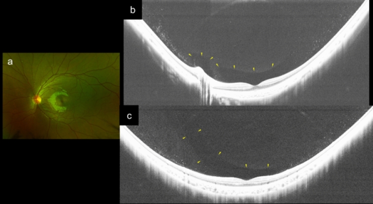
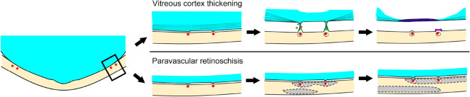
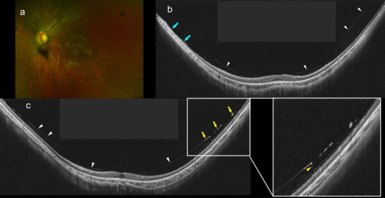
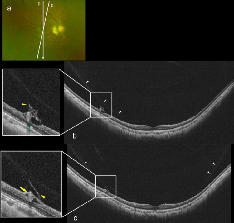
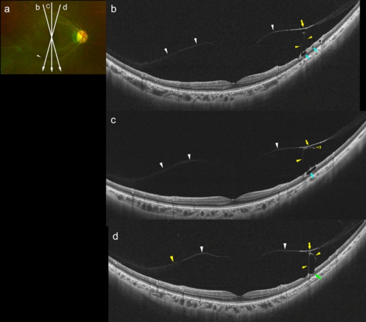
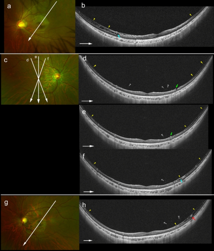
The purpose of this study was to determine the relationship between a posterior vitreous detachment (PVD) and retinoschisis (RS) in 73 highly myopic (HM) young patients age 16.4 ± 6.9 years and 24 non-HM children age 8.4 ± 1.5 years. The presence of the paravascular retinal abnormalities was determined in the images obtained by a ultra-widefield OCT (UWF OCT) instrument with an image field of 23 × 20 mm. The results showed that a partial PVD was detected in 15 (21%) of the HM patients, and the number increased significantly with increasing age (P = 0.02). PVDs of any type were not found in the non-HM eyes. The number of microvascular folds also increased with age in the HM patients (P = 0.03). Medium-reflective columnar tissues were present between the detached vitreous and inner retinal surface in 4 (5%) eyes of the HM patients. Myopic RS was found in 3 (4%) HM patients in the paravascular area but not in the macular area. These results suggest that early partial PVD may play a role in pathological and proliferative vitreous changes of HM eyes. An intense vitreoretinal traction with bridging tissues may cause the various paravascular retinal abnormalities. In HM eyes, paravascular RS is already present at an early age which may progress to macular RS with aging.

摘要

本研究旨在探讨 73 例高度近视(HM)青年患者(年龄 16.4±6.9 岁)和 24 例非 HM 儿童(年龄 8.4±1.5 岁)的后玻璃体脱离(PVD)与格子样视网膜劈裂(RS)之间的关系。通过超宽视野 OCT(UWF OCT)仪器获取的图像,确定视网膜旁血管异常的存在,该仪器的图像视野为 23×20mm。结果显示,15 例(21%)HM 患者存在部分 PVD,且随年龄增长而显著增加(P=0.02)。非 HM 眼未发现任何类型的 PVD。HM 患者的微血管褶皱数量也随年龄增长而增加(P=0.03)。在 4 例(5%)HM 患者的眼中,脱离的玻璃体和内视网膜表面之间存在中等反射柱状组织。HM 患者的血管旁区域发现 3 例(4%)近视性 RS,但在黄斑区域未发现。这些结果表明,早期的部分 PVD 可能在 HM 眼的病理性和增殖性玻璃体改变中起作用。强烈的玻璃体视网膜牵引和桥接组织可能导致各种血管旁视网膜异常。在 HM 眼中,血管旁 RS 早在年轻时就已经存在,随着年龄的增长可能会进展为黄斑 RS。

https://cdn.ncbi.nlm.nih.gov/pmc/blobs/2fd0/8405667/3b599fc24cd6/41598_2021_96783_Fig7_HTML.jpg
https://cdn.ncbi.nlm.nih.gov/pmc/blobs/2fd0/8405667/f9ca84eef6d0/41598_2021_96783_Fig1_HTML.jpg
https://cdn.ncbi.nlm.nih.gov/pmc/blobs/2fd0/8405667/6fe37790b59e/41598_2021_96783_Fig2_HTML.jpg
https://cdn.ncbi.nlm.nih.gov/pmc/blobs/2fd0/8405667/e8dc6afcf61b/41598_2021_96783_Fig3_HTML.jpg
https://cdn.ncbi.nlm.nih.gov/pmc/blobs/2fd0/8405667/456454710ef9/41598_2021_96783_Fig4_HTML.jpg
https://cdn.ncbi.nlm.nih.gov/pmc/blobs/2fd0/8405667/336a764bd2bd/41598_2021_96783_Fig5_HTML.jpg
https://cdn.ncbi.nlm.nih.gov/pmc/blobs/2fd0/8405667/b83e9a68e58e/41598_2021_96783_Fig6_HTML.jpg
https://cdn.ncbi.nlm.nih.gov/pmc/blobs/2fd0/8405667/3b599fc24cd6/41598_2021_96783_Fig7_HTML.jpg

相似文献

1
Posterior vitreous detachment and paravascular retinoschisis in highly myopic young patients detected by ultra-widefield OCT.高度近视年轻患者的超广角 OCT 检测到后玻璃体脱离和血管旁视网膜劈裂。
Sci Rep. 2021 Aug 30;11(1):17330. doi: 10.1038/s41598-021-96783-w.
2
Ultra-Widefield Optical Coherence Tomographic Imaging of Posterior Vitreous in Eyes With High Myopia.高度近视眼中的后玻璃体超广角光相干断层扫描成像。
Am J Ophthalmol. 2019 Oct;206:102-112. doi: 10.1016/j.ajo.2019.03.011. Epub 2019 Mar 20.
3
Importance of Paravascular Vitreal Adhesions for Development of Myopic Macular Retinoschisis Detected by Ultra-Widefield OCT.重要的是,血管旁玻璃体内粘连对于通过超广角 OCT 检测到的近视性黄斑视网膜劈裂的发展。
Ophthalmology. 2021 Feb;128(2):256-265. doi: 10.1016/j.ophtha.2020.06.063. Epub 2020 Jul 11.
4
Relationship Between Paravascular Abnormalities and Choroidal Thickness in Young Highly Myopic Adults.年轻人高度近视中脉络膜厚度与管周异常的关系。
Transl Vis Sci Technol. 2022 Jun 1;11(6):18. doi: 10.1167/tvst.11.6.18.
5
Detection of paravascular lamellar holes and other paravascular abnormalities by optical coherence tomography in eyes with high myopia.光学相干断层扫描检测高度近视眼中的血管旁板层孔及其他血管旁异常情况。
Ophthalmology. 2008 Apr;115(4):708-17. doi: 10.1016/j.ophtha.2007.04.060. Epub 2007 Aug 29.
6
OBSERVATION OF VITREOUS FEATURES USING ENHANCED VITREOUS IMAGING OPTICAL COHERENCE TOMOGRAPHY IN HIGHLY MYOPIC RETINOSCHISIS.高度近视性视网膜劈裂症中应用改良玻璃体成像光相干断层扫描观察玻璃体特征。
Retina. 2019 Sep;39(9):1732-1741. doi: 10.1097/IAE.0000000000002226.
7
EVALUATION OF PARAVASCULAR INNER RETINAL DEFECTS USING EN FACE OPTICAL COHERENCE TOMOGRAPHY.应用光学相干断层扫描评估血管周围内层视网膜病变。
Retina. 2023 Oct 1;43(10):1644-1652. doi: 10.1097/IAE.0000000000003889.
8
Paravascular abnormalities observed by spectral domain optical coherence tomography are risk factors for retinoschisis in eyes with high myopia.频域光相干断层扫描观察到的脉络膜旁异常是高度近视患者发生格子样视网膜劈裂的危险因素。
Acta Ophthalmol. 2018 Jun;96(4):e515-e523. doi: 10.1111/aos.13628. Epub 2017 Nov 24.
9
Clinical characteristics and risk factors of myopic retinoschisis in an elderly high myopia population.老年高度近视人群中近视性视网膜劈裂的临床特征及危险因素。
Acta Ophthalmol. 2023 Mar;101(2):e167-e176. doi: 10.1111/aos.15234. Epub 2022 Aug 25.
10
[Clinical observations of macular hole with and without retinal detachment in high myopic eyes].高度近视眼黄斑裂孔伴或不伴视网膜脱离的临床观察
Zhonghua Yan Ke Za Zhi. 2010 Jun;46(6):508-12.

引用本文的文献

1
Posterior Vitreous Detachment in Healthy Versus AMD Eyes Assessed by Widefield Optical Coherence Tomography.通过广角光学相干断层扫描评估健康眼与年龄相关性黄斑变性(AMD)眼的玻璃体后脱离情况
Diagnostics (Basel). 2025 May 29;15(11):1382. doi: 10.3390/diagnostics15111382.
2
Silicone oil emulsification: A literature review and role of widefield imaging and ultra-widefield imaging with navigated central and peripheral optical coherence tomography technology.硅油乳化:文献综述以及宽视野成像和导航式中心及周边光学相干断层扫描技术的超广角成像的作用
Saudi J Ophthalmol. 2024 Jan 6;38(2):112-122. doi: 10.4103/sjopt.sjopt_193_23. eCollection 2024 Apr-Jun.
3

本文引用的文献

1
Importance of Paravascular Vitreal Adhesions for Development of Myopic Macular Retinoschisis Detected by Ultra-Widefield OCT.重要的是,血管旁玻璃体内粘连对于通过超广角 OCT 检测到的近视性黄斑视网膜劈裂的发展。
Ophthalmology. 2021 Feb;128(2):256-265. doi: 10.1016/j.ophtha.2020.06.063. Epub 2020 Jul 11.
2
Comparison of Vitreomacular Interface Changes in Myopic Foveoschisis and Idiopathic Epiretinal Membrane Foveoschisis.近视性黄斑劈裂与特发性视网膜内界膜黄斑劈裂的玻璃体黄斑界面改变比较。
Am J Ophthalmol. 2020 Sep;217:152-161. doi: 10.1016/j.ajo.2020.04.023. Epub 2020 Apr 29.
3
Posterior Vitreous Detachment in Highly Myopic Patients.
Multimodal imaging of optic nerve head abnormalities in high myopia.
高度近视性视神经乳头异常的多模态成像
Front Neurol. 2024 Apr 23;15:1366593. doi: 10.3389/fneur.2024.1366593. eCollection 2024.
4
Posterior Vitreous Detachment and Its Role in the Evolution of Dry to Wet Age Related Macular Degeneration [Letter].玻璃体后脱离及其在干性至湿性年龄相关性黄斑变性演变中的作用[信函]
Clin Ophthalmol. 2023 Mar 27;17:991-992. doi: 10.2147/OPTH.S413232. eCollection 2023.
5
Machine learning-based 3D modeling and volumetry of human posterior vitreous cavity of optical coherence tomographic images.基于机器学习的光学相干断层扫描图像人眼后玻璃体腔的 3D 建模与容积测量。
Sci Rep. 2022 Aug 16;12(1):13836. doi: 10.1038/s41598-022-17615-z.
6
Progress of Imaging in Diabetic Retinopathy-From the Past to the Present.糖尿病视网膜病变的影像学进展——从过去到现在
Diagnostics (Basel). 2022 Jul 11;12(7):1684. doi: 10.3390/diagnostics12071684.
7
Relationship Between Paravascular Abnormalities and Choroidal Thickness in Young Highly Myopic Adults.年轻人高度近视中脉络膜厚度与管周异常的关系。
Transl Vis Sci Technol. 2022 Jun 1;11(6):18. doi: 10.1167/tvst.11.6.18.
高度近视患者的玻璃体后脱离。
Invest Ophthalmol Vis Sci. 2020 Apr 9;61(4):33. doi: 10.1167/iovs.61.4.33.
4
Ultra-Widefield Optical Coherence Tomographic Imaging of Posterior Vitreous in Eyes With High Myopia.高度近视眼中的后玻璃体超广角光相干断层扫描成像。
Am J Ophthalmol. 2019 Oct;206:102-112. doi: 10.1016/j.ajo.2019.03.011. Epub 2019 Mar 20.
5
Posterior Vitreous Detachment as Observed by Wide-Angle OCT Imaging.宽角 OCT 成像观察到的后玻璃体脱离。
Ophthalmology. 2018 Sep;125(9):1372-1383. doi: 10.1016/j.ophtha.2018.02.039. Epub 2018 Apr 7.
6
Posterior Staphylomas in Pathologic Myopia Imaged by Widefield Optical Coherence Tomography.广角光学相干断层扫描成像的病理性近视后巩膜葡萄肿
Invest Ophthalmol Vis Sci. 2017 Jul 1;58(9):3750-3758. doi: 10.1167/iovs.17-22319.
7
Application of extended field imaging to optical coherence tomography.扩展视野成像在光学相干断层扫描中的应用。
Ophthalmology. 2015 Jun;122(6):1272-4. doi: 10.1016/j.ophtha.2014.12.035. Epub 2015 Jan 29.
8
Vitreous changes in high myopia observed by swept-source optical coherence tomography.扫频源光学相干断层扫描观察高度近视的玻璃体改变。
Invest Ophthalmol Vis Sci. 2014 Mar 10;55(3):1447-52. doi: 10.1167/iovs.13-13496.
9
The diversity of traction mechanisms in myopic traction maculopathy.近视牵引性脉络膜病变中牵引机制的多样性。
Am J Ophthalmol. 2012 Jan;153(1):93-102. doi: 10.1016/j.ajo.2011.06.016. Epub 2011 Sep 9.
10
Observations by spectral-domain optical coherence tomography combined with simultaneous scanning laser ophthalmoscopy: imaging of the vitreous.频域光相干断层扫描结合同步扫描激光检眼镜观察:玻璃体成像。
Am J Ophthalmol. 2010 Apr;149(4):641-50. doi: 10.1016/j.ajo.2009.11.016. Epub 2010 Feb 9.