• 文献检索
  • 文档翻译
  • 深度研究
  • 学术资讯
  • Suppr Zotero 插件Zotero 插件
  • 邀请有礼
  • 套餐&价格
  • 历史记录
应用&插件
Suppr Zotero 插件Zotero 插件浏览器插件Mac 客户端Windows 客户端微信小程序
定价
高级版会员购买积分包购买API积分包
服务
文献检索文档翻译深度研究API 文档MCP 服务
关于我们
关于 Suppr公司介绍联系我们用户协议隐私条款
关注我们

Suppr 超能文献

核心技术专利:CN118964589B侵权必究
粤ICP备2023148730 号-1Suppr @ 2026

文献检索

告别复杂PubMed语法,用中文像聊天一样搜索,搜遍4000万医学文献。AI智能推荐,让科研检索更轻松。

立即免费搜索

文件翻译

保留排版,准确专业,支持PDF/Word/PPT等文件格式,支持 12+语言互译。

免费翻译文档

深度研究

AI帮你快速写综述,25分钟生成高质量综述,智能提取关键信息,辅助科研写作。

立即免费体验

脑部MRI诊断特发性颅内高压的定量参数

Quantitative parameters for diagnosis of idiopathic intracranial hypertension on brain MRI.

作者信息

Marashdeh Wael M, Al Qaralleh Mohammad A, Hdeeb Ahmad H

机构信息

Jordan University of Science and Technology, King Abdullah University Hospital, Department of Diagnostic Radiology and Nuclear Medicine, Jordan.

出版信息

Eur J Radiol Open. 2021 Aug 26;8:100371. doi: 10.1016/j.ejro.2021.100371. eCollection 2021.

DOI:10.1016/j.ejro.2021.100371
PMID:34485627
原文链接:https://pmc.ncbi.nlm.nih.gov/articles/PMC8403732/
Abstract

PURPOSE

Aim of this study was to develop quantitative parameters for diagnosing Idiopathic Intracranial Hypertension (IIH) using brain MRI scans.

METHODS

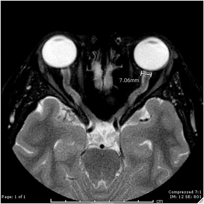
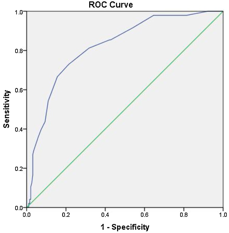
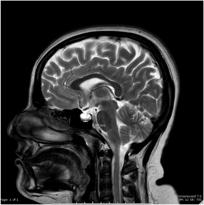
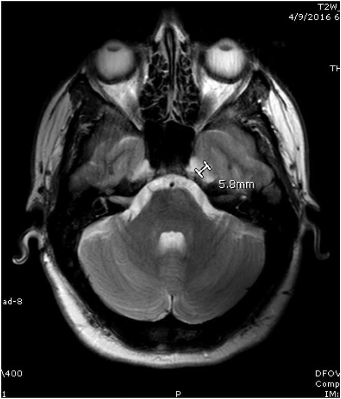
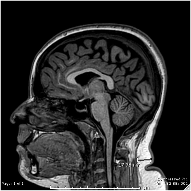
This is a case control study with 48 cases and 192 matched controls. Optic nerve diameter (OND), Pituitary height (PH), Meckel's cave diameter (MCD), and Neck fat thickness (NFT) were measured for both groups. Consequently, means were obtained for the different parameters in both groups with subsequent establishment of best cutoffs using Receiver Operator Curve (ROC) analysis.

RESULTS

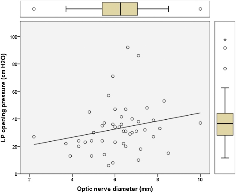
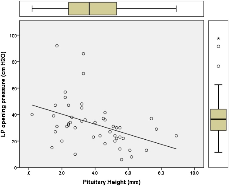
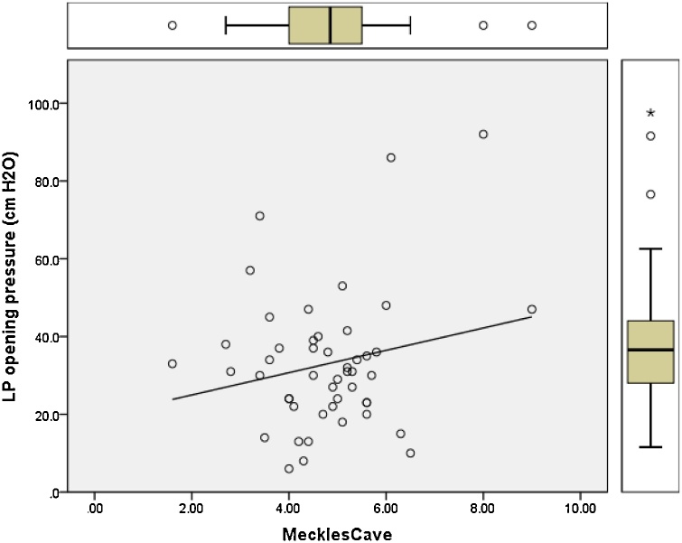
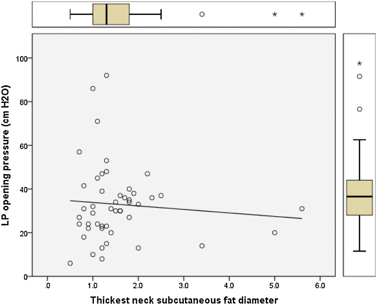
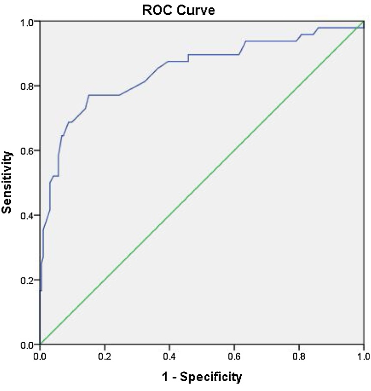
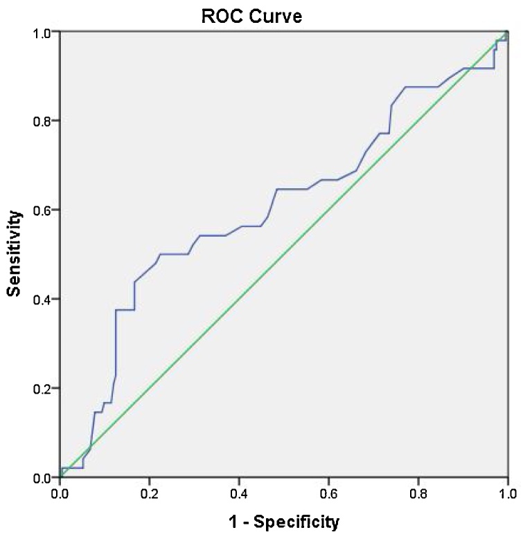
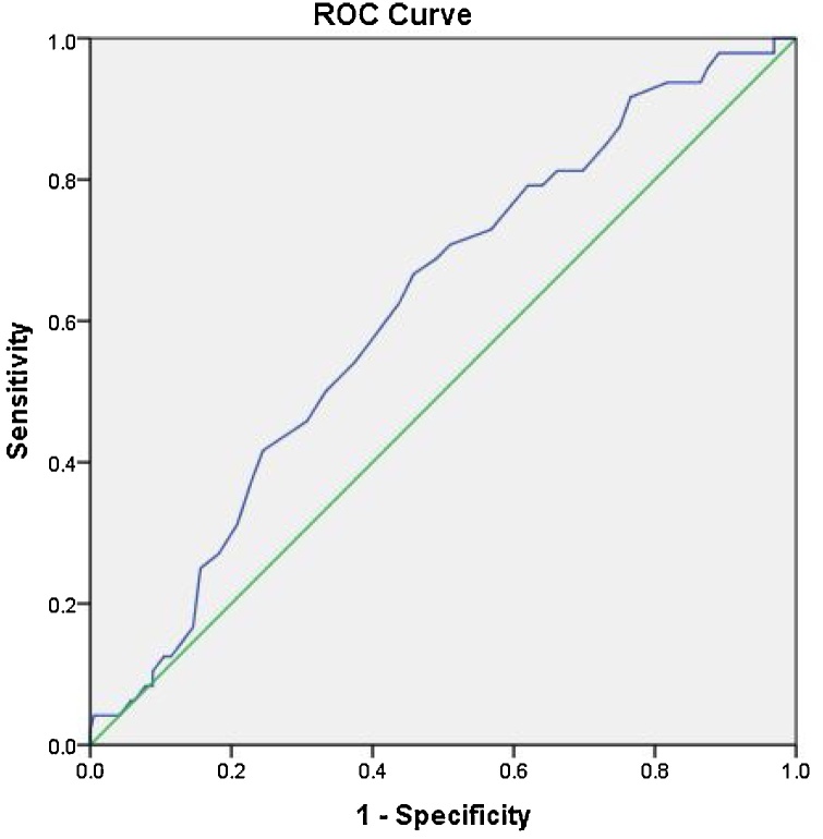
For IIH patients the means of OND, PH, MCD, and NFT were 6.2 mm, 3.9 mm, 5 mm, 1.4 cm, respectively while for controls the means were 4.6 mm, 4.5 mm, 4.3 mm, and 0.8 cm with statistical significance between the two groups. ROC analysis showed the cutoff points with best accuracy for the above parameters in diagnosing IIH to be 5.4 mm for OND with sensitivity of 0.77 and specificity of 0.85 representing high accuracy, while for PH a cutoff point of 3 mm showed low accuracy with sensitivity of 0.54 and specificity of 0.7, and a MCD cutoff of 4.5 mm also showed low accuracy with sensitivity of 0.6 and specificity of 0.59, meanwhile a cutoff point of 1.1 cm for NFT was moderately accurate with sensitivity of 0.70 and specificity of 0.81.

CONCLUSION

Statistical difference in the means for OND, PH, MCD, and NFT between IIH patients and controls is established. Also, we provide cut off points for these parameters to diagnose IIH on brain MRI.

摘要

目的

本研究的目的是利用脑部磁共振成像(MRI)扫描开发诊断特发性颅内高压(IIH)的定量参数。

方法

这是一项病例对照研究,有48例病例和192例匹配对照。对两组均测量了视神经直径(OND)、垂体高度(PH)、 Meckel腔直径(MCD)和颈部脂肪厚度(NFT)。随后,获得两组不同参数的均值,并使用受试者操作特征曲线(ROC)分析确定最佳截断值。

结果

IIH患者的OND、PH、MCD和NFT均值分别为6.2毫米、3.9毫米、5毫米、1.4厘米,而对照组的均值分别为4.6毫米、4.5毫米、4.3毫米和0.8厘米,两组之间具有统计学意义。ROC分析显示,上述参数在诊断IIH时具有最佳准确性的截断点,OND为5.4毫米,灵敏度为0.77,特异性为0.85,代表高准确性;而PH的截断点为3毫米,准确性较低,灵敏度为0.54,特异性为0.7;MCD的截断点为4.5毫米,准确性也较低,灵敏度为0.6,特异性为0.59;同时,NFT的截断点为1.1厘米,准确性中等,灵敏度为0.70,特异性为0.81。

结论

IIH患者与对照组在OND、PH、MCD和NFT均值上存在统计学差异。此外,我们提供了这些参数在脑部MRI上诊断IIH的截断点。

https://cdn.ncbi.nlm.nih.gov/pmc/blobs/b35c/8403732/3ebe9e3e6d92/gr12.jpg
https://cdn.ncbi.nlm.nih.gov/pmc/blobs/b35c/8403732/de87a98c0f9a/gr1.jpg
https://cdn.ncbi.nlm.nih.gov/pmc/blobs/b35c/8403732/35c5fce3b498/gr2.jpg
https://cdn.ncbi.nlm.nih.gov/pmc/blobs/b35c/8403732/90378423b45d/gr3.jpg
https://cdn.ncbi.nlm.nih.gov/pmc/blobs/b35c/8403732/02941b5584be/gr4.jpg
https://cdn.ncbi.nlm.nih.gov/pmc/blobs/b35c/8403732/e439e5f0128c/gr5.jpg
https://cdn.ncbi.nlm.nih.gov/pmc/blobs/b35c/8403732/2065ef9eb2d1/gr6.jpg
https://cdn.ncbi.nlm.nih.gov/pmc/blobs/b35c/8403732/3ae977255ed8/gr7.jpg
https://cdn.ncbi.nlm.nih.gov/pmc/blobs/b35c/8403732/1b8d71b5d3e6/gr8.jpg
https://cdn.ncbi.nlm.nih.gov/pmc/blobs/b35c/8403732/d3b9758c6d74/gr9.jpg
https://cdn.ncbi.nlm.nih.gov/pmc/blobs/b35c/8403732/3a34feac79f5/gr10.jpg
https://cdn.ncbi.nlm.nih.gov/pmc/blobs/b35c/8403732/f8080a3041a8/gr11.jpg
https://cdn.ncbi.nlm.nih.gov/pmc/blobs/b35c/8403732/3ebe9e3e6d92/gr12.jpg

相似文献

1
Quantitative parameters for diagnosis of idiopathic intracranial hypertension on brain MRI.脑部MRI诊断特发性颅内高压的定量参数
Eur J Radiol Open. 2021 Aug 26;8:100371. doi: 10.1016/j.ejro.2021.100371. eCollection 2021.
2
Noticable Findings in Cranial MRI of the Patients with Idiopathic Intracranial Hypertension.特发性颅内高压患者头颅磁共振成像的显著发现。
Am J Rhinol Allergy. 2022 Jul;36(4):415-422. doi: 10.1177/19458924211069213. Epub 2022 Mar 2.
3
Optic nerve sonographic examination to predict raised intracranial pressure in idiopathic intracranial hypertension: The cut-off points.视神经超声检查预测特发性颅内高压患者颅内压升高:切点分析
Neuroradiol J. 2018 Oct;31(5):490-495. doi: 10.1177/1971400918789385. Epub 2018 Jul 19.
4
Sonographic assessment of optic nerve and ophthalmic vessels in patients with idiopathic intracranial hypertension.特发性颅内高压患者视神经和眼血管的超声评估
Neurol Res. 2018 Sep;40(9):728-735. doi: 10.1080/01616412.2018.1473097. Epub 2018 May 25.
5
Utility of Magnetic Resonance Imaging Features for Improving the Diagnosis of Idiopathic Intracranial Hypertension Without Papilledema.磁共振成像特征在提高无视乳头水肿特发性颅内高压症诊断中的作用。
J Neuroophthalmol. 2019 Sep;39(3):299-307. doi: 10.1097/WNO.0000000000000767.
6
Quantifying response to intracranial pressure normalization in idiopathic intracranial hypertension via dynamic neuroimaging.通过动态神经影像学量化特发性颅内高压患者颅内压正常化的反应。
J Magn Reson Imaging. 2018 Apr;47(4):913-927. doi: 10.1002/jmri.25857. Epub 2017 Sep 27.
7
Combined accuracy of optic nerve sheath diameter, strain ratio, and shear wave elastography of the optic nerve in patients with idiopathic intracranial hypertension.特发性颅内高压患者视神经鞘直径、应变率及视神经剪切波弹性成像的联合准确性
Ultrasonography. 2022 Jan;41(1):106-113. doi: 10.14366/usg.20165. Epub 2021 Apr 20.
8
MR findings suggestive of idiopathic intracranial hypertension in 117 patients with spontaneous cerebrospinal fluid rhinorrhea.117例自发性脑脊液鼻漏患者的磁共振成像(MR)表现提示特发性颅内高压。
Neuroradiology. 2022 May;64(5):949-958. doi: 10.1007/s00234-021-02840-6. Epub 2021 Oct 22.
9
Sonographic assessment of the optic nerve sheath in idiopathic intracranial hypertension.特发性颅内高压视神经鞘的超声评估。
J Neurol. 2011 Nov;258(11):2014-9. doi: 10.1007/s00415-011-6059-0. Epub 2011 Apr 28.
10
The significance of ONSD, ONSD/ETD ratio, and other neuroimaging parameters in idiopathic intracranial hypertension.颅内压增高症中 ONSD、ONSD/ETD 比值及其他神经影像学参数的意义。
Neurol Res. 2021 Dec;43(12):1098-1106. doi: 10.1080/01616412.2021.1949688. Epub 2021 Aug 19.

引用本文的文献

1
How can MRI descriptors be optimally combined to predict idiopathic intracranial hypertension?磁共振成像(MRI)描述符如何才能得到最佳组合以预测特发性颅内高压?
Br J Radiol. 2025 Jul 1;98(1171):1070-1079. doi: 10.1093/bjr/tqaf080.
2
Magnetic resonance signs of intracranial hypertension in children: a retrospective case-control study.儿童颅内高压的磁共振征象:一项回顾性病例对照研究。
Eur J Pediatr. 2025 Mar 1;184(3):218. doi: 10.1007/s00431-025-06025-8.
3
Idiopathic intracranial hypertension: Imaging and clinical fundamentals.特发性颅内高压:影像学与临床基础

本文引用的文献

1
High-resolution MRI assessment of optic nerve sheath diameter in adults: optic nerve sheath variation and a new diagnostic tool for intracranial hypertension.成人视神经鞘直径的高分辨率MRI评估:视神经鞘变异及颅内高压的一种新诊断工具。
Acta Radiol. 2021 Oct;62(10):1397-1403. doi: 10.1177/0284185120966715. Epub 2020 Oct 21.
2
A systematic review of surgical treatments of idiopathic intracranial hypertension (IIH).特发性颅内高压(IIH)的手术治疗的系统评价。
Neurosurg Rev. 2021 Apr;44(2):773-792. doi: 10.1007/s10143-020-01288-1. Epub 2020 Apr 25.
3
Idiopathic intracranial hypertension.
World J Radiol. 2024 Dec 28;16(12):722-748. doi: 10.4329/wjr.v16.i12.722.
4
Debilitating trigeminal neuralgia secondary to idiopathic intracranial hypertension.继发于特发性颅内高压的致残性三叉神经痛。
Heliyon. 2023 Sep 10;9(9):e19756. doi: 10.1016/j.heliyon.2023.e19756. eCollection 2023 Sep.
5
A rare finding of bilateral facial canal meningoceles involving the tympanic segment in suspected idiopathic intracranial hypertension.在疑似特发性颅内高压患者中罕见发现双侧面神经管脑膜膨出累及鼓室段。
Radiol Case Rep. 2023 Jun 28;18(9):3184-3187. doi: 10.1016/j.radcr.2023.06.036. eCollection 2023 Sep.
6
How to understand an enlarged Meckel's cave? An anatomical study.如何理解扩大的 Meckel 氏腔?一项解剖学研究。
Surg Radiol Anat. 2023 Aug;45(8):933-937. doi: 10.1007/s00276-023-03177-w. Epub 2023 Jun 15.
7
Efficacy of Dural Sinus Quantitative Measurements in Idiopathic Intracranial Hypertension : A Practical Diagnostic Feature.特发性颅内高压中硬脑膜窦定量测量的疗效:一种实用的诊断特征。
Clin Neuroradiol. 2023 Jun;33(2):545-554. doi: 10.1007/s00062-022-01244-0. Epub 2022 Dec 28.
特发性颅内高压
JAAPA. 2019 May;32(5):30-35. doi: 10.1097/01.JAA.0000554732.85914.91.
4
Measurement of the Optic Nerve Sheath Diameter with Magnetic Resonance Imaging and Its Association with Eyeball Diameter in Healthy Adults.健康成年人中磁共振成像测量视神经鞘直径及其与眼球直径的关系
J Clin Neurol. 2018 Jul;14(3):345-350. doi: 10.3988/jcn.2018.14.3.345. Epub 2018 May 31.
5
Quantitative Phase-Contrast MR Angiography to Measure Hemodynamic Changes in Idiopathic Intracranial Hypertension.定量相位对比磁共振血管成像测量特发性颅内高压的血流动力学变化。
AJNR Am J Neuroradiol. 2018 Apr;39(4):682-686. doi: 10.3174/ajnr.A5571. Epub 2018 Mar 8.
6
Transverse Sinus Stenosis Is the Most Sensitive MR Imaging Correlate of Idiopathic Intracranial Hypertension.横窦狭窄是特发性颅内高压最敏感的磁共振成像相关表现。
AJNR Am J Neuroradiol. 2017 Mar;38(3):471-477. doi: 10.3174/ajnr.A5055. Epub 2017 Jan 19.
7
Imaging of Intracranial Pressure Disorders.颅内压紊乱的影像学检查
Neurosurgery. 2017 Mar 1;80(3):341-354. doi: 10.1227/NEU.0000000000001362.
8
Brain Imaging in Idiopathic Intracranial Hypertension.特发性颅内高压的脑成像
J Neuroophthalmol. 2015 Dec;35(4):400-11. doi: 10.1097/WNO.0000000000000303.
9
Association of MRI findings and visual outcome in idiopathic intracranial hypertension.特发性颅内高压的 MRI 表现与视觉预后的相关性。
AJR Am J Roentgenol. 2013 Aug;201(2):412-8. doi: 10.2214/AJR.12.9638.
10
Factors determining the clinical significance of an "empty" sella turcica.影响空蝶鞍临床意义的因素。
AJR Am J Roentgenol. 2013 May;200(5):1125-31. doi: 10.2214/AJR.12.9013.