Department of Anatomy and Embryology, Leiden University Medical Center, Leiden, The Netherlands.
Sequencing Analysis Support Core, Department of Biomedical Data Sciences, Leiden University Medical Center, Leiden, The Netherlands.
PLoS Genet. 2021 Sep 9;17(9):e1009773. doi: 10.1371/journal.pgen.1009773. eCollection 2021 Sep.
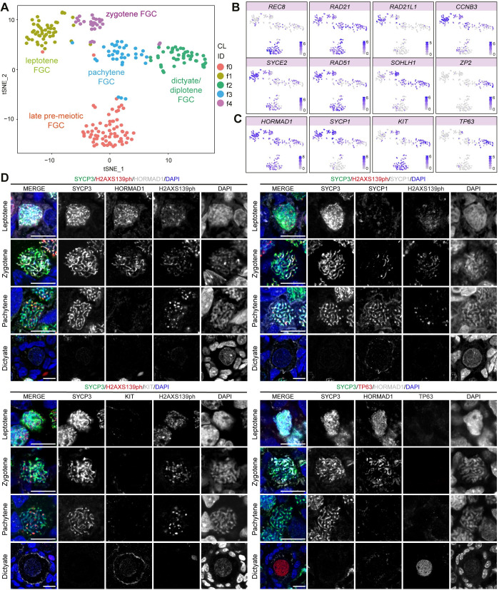
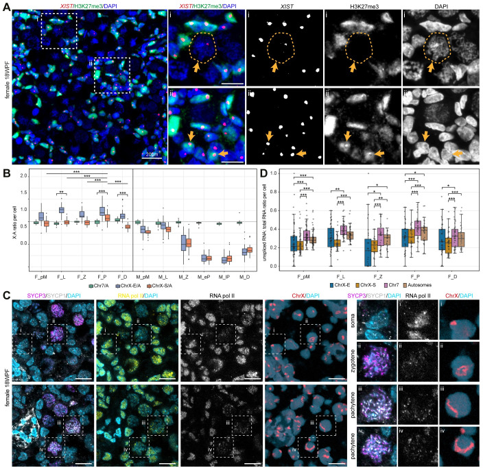
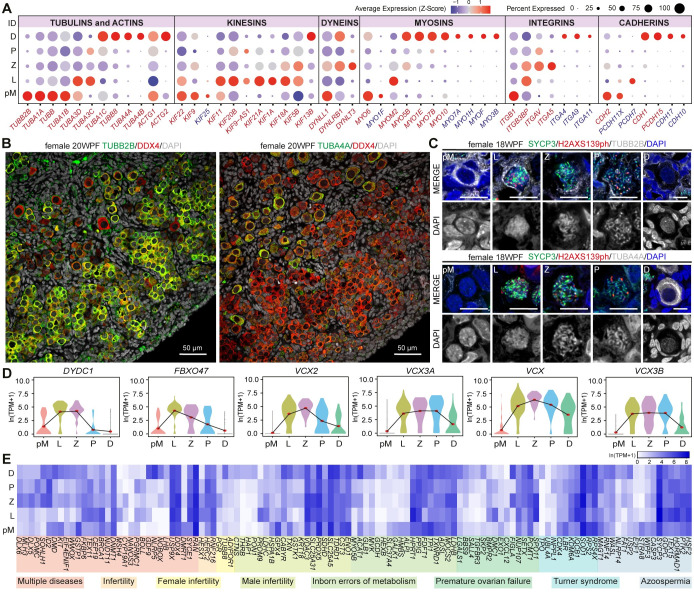
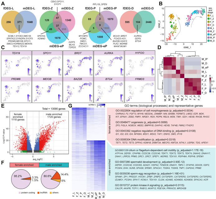
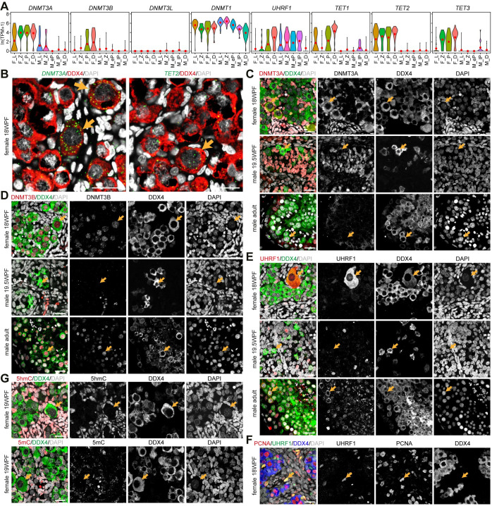
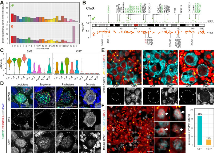
During gametogenesis in mammals, meiosis ensures the production of haploid gametes. The timing and length of meiosis to produce female and male gametes differ considerably. In contrast to males, meiotic prophase I in females initiates during development. Hence, the knowledge regarding progression through meiotic prophase I is mainly focused on human male spermatogenesis and female oocyte maturation during adulthood. Therefore, it remains unclear how the different stages of meiotic prophase I between human oogenesis and spermatogenesis compare. Analysis of single-cell transcriptomics data from human fetal germ cells (FGC) allowed us to identify the molecular signatures of female meiotic prophase I stages leptotene, zygotene, pachytene and diplotene. We have compared those between male and female germ cells in similar stages of meiotic prophase I and revealed conserved and specific features between sexes. We identified not only key players involved in the process of meiosis, but also highlighted the molecular components that could be responsible for changes in cellular morphology that occur during this developmental period, when the female FGC acquire their typical (sex-specific) oocyte shape as well as sex-differences in the regulation of DNA methylation. Analysis of X-linked expression between sexes during meiotic prophase I suggested a transient X-linked enrichment during female pachytene, that contrasts with the meiotic sex chromosome inactivation in males. Our study of the events that take place during meiotic prophase I provide a better understanding not only of female meiosis during development, but also highlights biomarkers that can be used to study infertility and offers insights in germline sex dimorphism in humans.
在哺乳动物的配子发生过程中,减数分裂确保了单倍体配子的产生。产生雌性和雄性配子的减数分裂的时间和长度有很大的不同。与男性不同,女性减数分裂前期 I 是在发育过程中开始的。因此,关于减数分裂前期 I 进展的知识主要集中在人类男性精子发生和成年女性卵母细胞成熟上。因此,尚不清楚人类卵发生和精子发生中减数分裂前期 I 的不同阶段如何比较。对人类胎儿生殖细胞 (FGC) 的单细胞转录组学数据的分析,使我们能够鉴定出减数分裂前期 I 的雌性细线期、合线期、粗线期和双线期的分子特征。我们将这些特征与减数分裂前期 I 相似阶段的雄性和雌性生殖细胞进行了比较,揭示了两性之间的保守和特定特征。我们不仅鉴定了参与减数分裂过程的关键因子,还强调了可能导致在这个发育时期细胞形态发生变化的分子成分,在此期间,女性 FGC 获得其典型的(性别特异性)卵母细胞形状以及 DNA 甲基化调控中的性别差异。对减数分裂前期 I 期间性染色体表达的分析表明,女性粗线期存在短暂的 X 连锁富集,与男性减数分裂性染色体失活形成对比。我们对减数分裂前期 I 期间发生的事件的研究,不仅更好地理解了发育过程中的雌性减数分裂,还强调了可以用于研究不孕不育的生物标志物,并为人类生殖细胞性别二态性提供了新的认识。