• 文献检索
  • 文档翻译
  • 深度研究
  • 学术资讯
  • Suppr Zotero 插件Zotero 插件
  • 邀请有礼
  • 套餐&价格
  • 历史记录
应用&插件
Suppr Zotero 插件Zotero 插件浏览器插件Mac 客户端Windows 客户端微信小程序
定价
高级版会员购买积分包购买API积分包
服务
文献检索文档翻译深度研究API 文档MCP 服务
关于我们
关于 Suppr公司介绍联系我们用户协议隐私条款
关注我们

Suppr 超能文献

核心技术专利:CN118964589B侵权必究
粤ICP备2023148730 号-1Suppr @ 2026

文献检索

告别复杂PubMed语法,用中文像聊天一样搜索,搜遍4000万医学文献。AI智能推荐,让科研检索更轻松。

立即免费搜索

文件翻译

保留排版,准确专业,支持PDF/Word/PPT等文件格式,支持 12+语言互译。

免费翻译文档

深度研究

AI帮你快速写综述,25分钟生成高质量综述,智能提取关键信息,辅助科研写作。

立即免费体验

多光子显微镜与质谱联用揭示肝细胞代谢异质性

Multiphoton Microscopy and Mass Spectrometry for Revealing Metabolic Heterogeneity of Hepatocytes .

机构信息

Junior Researcher, Laboratory of Regenerative Medicine, Research Institute of Experimental Oncology and Biomedical Technologies, Privolzhsky Research Medical University, 10/1 Minin and Pozharsky Square, Nizhny Novgorod, 603005, Russia; PhD Student, Institute of Biology and Biomedicine, National Research Lobachevsky State University of Nizhni Novgorod, 23 Prospekt Gagarina, Nizhny Novgorod, 603950, Russia.

Researcher, Laboratory of Regenerative Medicine, Research Institute of Experimental Oncology and Biomedical Technologies, Privolzhsky Research Medical University, 10/1 Minin and Pozharsky Square, Nizhny Novgorod, 603005, Russia.

出版信息

Sovrem Tekhnologii Med. 2021;13(2):18-29. doi: 10.17691/stm2021.13.2.02. Epub 2021 Apr 30.

DOI:10.17691/stm2021.13.2.02
PMID:34513073
原文链接:https://pmc.ncbi.nlm.nih.gov/articles/PMC8353720/
Abstract

UNLABELLED

was to study the possibility of revealing the heterogeneity of normal liver hepatocytes in terms of metabolic status using the modern methods of multiphoton microscopy and mass spectrometry.

MATERIALS AND METHODS

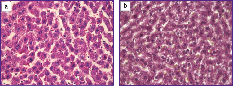
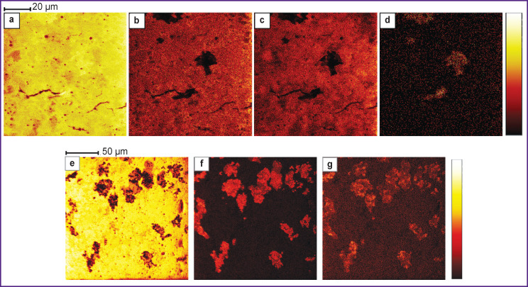
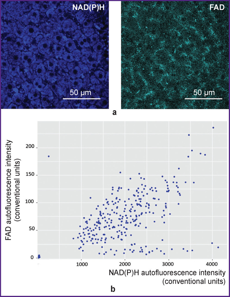
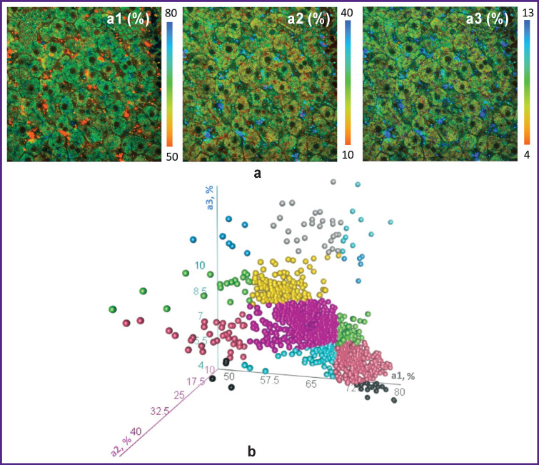
Heterogeneity of hepatocytes in terms of total metabolic activity was assessed using multiphoton microscopy based on the autofluorescence intensity of intracellular cofactors NAD(P)H and FAD. Hepatocyte heterogeneity in terms of intensity of intracellular metabolic processes was determined using the fluorescence lifetime imaging (FLIM) method based on the data about fluorescence lifetime contributions of various forms of NAD(P)H. The method of time-of-flight secondary ion mass spectrometry (TоF-SIMS) was used to study the lipid and amino acid composition of hepatocytes.

RESULTS

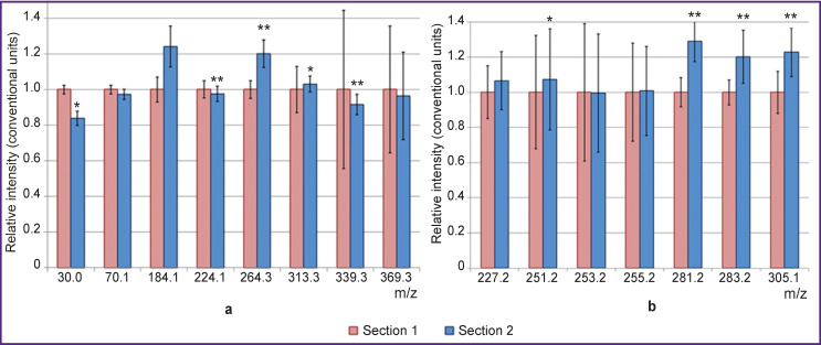
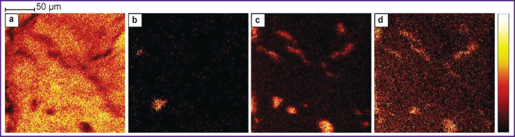
It has been revealed using multiphoton microscopy that hepatocytes are heterogeneous in terms of general metabolic activity. Using FLIM, it was established that the heterogeneity degree was high in terms of intensity of oxidative phosphorylation, glycolysis, and synthetic processes (lipogenesis, nucleic acid synthesis, and the pentose phosphate pathway). The TоF-SIMS method revealed the presence of hepatocyte heterogeneity in terms of amino acid and lipid composition, which points to various intensities of synthetic processes in individual hepatocytes. Moreover, differences in the content of PO ions were revealed. The results of ToF-SIMS study correlate with the data obtained by multiphoton microscopy and FLIM, confirming the revealed heterogeneity of hepatocytes in terms of general metabolic activity and intensity of intercellular metabolic processes.

CONCLUSION

The latest methods of fluorescence bioimaging and mass spectrometry proved to be effective in revealing hepatocyte heterogeneity in terms of metabolic status. The presence of heterogeneity should be taken into account in studying the liver tissue under various conditions with the application of fluorescence bioimaging methods.

摘要

目的

使用多光子显微镜和质谱等现代方法,研究正常肝细胞在代谢状态方面表现出异质性的可能性。

材料与方法

使用基于细胞内辅因子 NAD(P)H 和 FAD 自发荧光强度的多光子显微镜评估肝细胞在总代谢活性方面的异质性。使用基于各种形式 NAD(P)H 荧光寿命贡献数据的荧光寿命成像 (FLIM) 方法确定肝细胞在细胞内代谢过程强度方面的异质性。采用飞行时间二次离子质谱法 (TоF-SIMS) 研究肝细胞的脂质和氨基酸组成。

结果

多光子显微镜显示,肝细胞在一般代谢活性方面存在异质性。FLIM 研究表明,氧化磷酸化、糖酵解和合成过程(脂肪生成、核酸合成和戊糖磷酸途径)的强度具有高度的异质性程度。TоF-SIMS 方法揭示了氨基酸和脂质组成方面的肝细胞异质性,表明个别肝细胞中合成过程的强度存在差异。此外,还发现了 PO 离子含量的差异。TоF-SIMS 研究的结果与多光子显微镜和 FLIM 获得的数据相关,证实了肝细胞在一般代谢活性和细胞间代谢过程强度方面存在异质性。

结论

荧光生物成像和质谱的最新方法在揭示代谢状态下肝细胞的异质性方面是有效的。在应用荧光生物成像方法研究各种条件下的肝组织时,应考虑到异质性的存在。

https://cdn.ncbi.nlm.nih.gov/pmc/blobs/d7cb/8353720/22454733f0ea/STM-13-2-02-f6.jpg
https://cdn.ncbi.nlm.nih.gov/pmc/blobs/d7cb/8353720/debd9a93c079/STM-13-2-02-f1.jpg
https://cdn.ncbi.nlm.nih.gov/pmc/blobs/d7cb/8353720/cb925053ad31/STM-13-2-02-f2.jpg
https://cdn.ncbi.nlm.nih.gov/pmc/blobs/d7cb/8353720/fed41ae9923c/STM-13-2-02-f3.jpg
https://cdn.ncbi.nlm.nih.gov/pmc/blobs/d7cb/8353720/7a5a4666147c/STM-13-2-02-f4.jpg
https://cdn.ncbi.nlm.nih.gov/pmc/blobs/d7cb/8353720/805df7fba444/STM-13-2-02-f5.jpg
https://cdn.ncbi.nlm.nih.gov/pmc/blobs/d7cb/8353720/22454733f0ea/STM-13-2-02-f6.jpg

相似文献

1
Multiphoton Microscopy and Mass Spectrometry for Revealing Metabolic Heterogeneity of Hepatocytes .多光子显微镜与质谱联用揭示肝细胞代谢异质性
Sovrem Tekhnologii Med. 2021;13(2):18-29. doi: 10.17691/stm2021.13.2.02. Epub 2021 Apr 30.
2
Two-photon FLIM of NAD(P)H and FAD in mesenchymal stem cells undergoing either osteogenic or chondrogenic differentiation.对正在经历成骨或软骨分化的间充质干细胞中烟酰胺腺嘌呤二核苷酸(磷酸)(NAD(P)H)和黄素腺嘌呤二核苷酸(FAD)进行双光子荧光寿命成像。
Stem Cell Res Ther. 2017 Jan 28;8(1):15. doi: 10.1186/s13287-017-0484-7.
3
Insights into Metabolic Activity and Structure of the Retina through Multiphoton Fluorescence Lifetime Imaging Microscopy in Mice.通过多光子荧光寿命成像显微镜观察小鼠视网膜的代谢活性和结构。
Cells. 2022 Jul 22;11(15):2265. doi: 10.3390/cells11152265.
4
Autofluorescence lifetime imaging of cellular metabolism: Sensitivity toward cell density, pH, intracellular, and intercellular heterogeneity.细胞代谢的自体荧光寿命成像:对细胞密度、pH 值、细胞内和细胞间异质性的敏感性。
Cytometry A. 2019 Jan;95(1):56-69. doi: 10.1002/cyto.a.23603. Epub 2018 Oct 8.
5
Metabolic imaging and secondary ion mass spectrometry to define the structure and function of liver with acute and chronic pathology.代谢成像和二次离子质谱分析,用于定义急性和慢性病理状态下肝脏的结构和功能。
J Biomed Opt. 2019 Dec;25(1):1-14. doi: 10.1117/1.JBO.25.1.014508.
6
Multiphoton FLIM imaging of NAD(P)H and FAD with one excitation wavelength.利用单一激发波长的多光子 FLIM 成像技术对 NAD(P)H 和 FAD 进行成像。
J Biomed Opt. 2020 Jan;25(1):1-16. doi: 10.1117/1.JBO.25.1.014510.
7
Two-photon fluorescence lifetime imaging of intrinsic NADH in three-dimensional tumor models.三维肿瘤模型中内源性 NADH 的双光子荧光寿命成像。
Cytometry A. 2019 Jan;95(1):80-92. doi: 10.1002/cyto.a.23632. Epub 2018 Oct 21.
8
The Effect of Diabetes Mellitus Type 1 on the Energy Metabolism of Hepatocytes: Multiphoton Microscopy and Fluorescence Lifetime Imaging.1 型糖尿病对肝细胞能量代谢的影响:多光子显微镜和荧光寿命成像。
Int J Mol Sci. 2023 Nov 30;24(23):17016. doi: 10.3390/ijms242317016.
9
Contribution of autofluorescence from intracellular proteins in multiphoton fluorescence lifetime imaging.细胞内蛋白的自发荧光对多光子荧光寿命成像的贡献。
Sci Rep. 2022 Oct 5;12(1):16584. doi: 10.1038/s41598-022-20857-6.
10
In vivo multiphoton fluorescence lifetime imaging of protein-bound and free nicotinamide adenine dinucleotide in normal and precancerous epithelia.正常上皮和癌前上皮中蛋白质结合型和游离烟酰胺腺嘌呤二核苷酸的体内多光子荧光寿命成像
J Biomed Opt. 2007 Mar-Apr;12(2):024014. doi: 10.1117/1.2717503.

引用本文的文献

1
FLIMB: fluorescence lifetime microendoscopy for metabolic and functional imaging of femoral marrow at subcellular resolution.FLIMB:用于亚细胞分辨率下股骨骨髓代谢和功能成像的荧光寿命显微内镜检查。
Biomed Opt Express. 2025 Mar 31;16(4):1711-1731. doi: 10.1364/BOE.549311. eCollection 2025 Apr 1.
2
Spatial metabolomics and its application in the liver.空间代谢组学及其在肝脏中的应用。
Hepatology. 2024 May 1;79(5):1158-1179. doi: 10.1097/HEP.0000000000000341. Epub 2023 Feb 23.
3
Multiplatform Metabolomics Studies of Human Cancers With NMR and Mass Spectrometry Imaging.

本文引用的文献

1
TOF-SIMS imaging reveals tumor heterogeneity and inflammatory response markers in the microenvironment of basal cell carcinoma.TOF-SIMS 成像揭示了基底细胞癌微环境中的肿瘤异质性和炎症反应标志物。
Biointerphases. 2020 Aug 28;15(4):041012. doi: 10.1116/6.0000340.
2
Multimodal Imaging Mass Spectrometry to Identify Markers of Pulmonary Arterial Hypertension in Human Lung Tissue Using MALDI-ToF, ToF-SIMS, and Hybrid SIMS.多模态成像质谱法结合 MALDI-TOF、TOF-SIMS 和杂交 SIMS 鉴定人肺组织肺动脉高压标志物
Anal Chem. 2020 Sep 1;92(17):12079-12087. doi: 10.1021/acs.analchem.0c02815. Epub 2020 Aug 17.
3
Resolving Metabolic Heterogeneity in Experimental Models of the Tumor Microenvironment from a Stable Isotope Resolved Metabolomics Perspective.
利用核磁共振和质谱成像技术对人类癌症进行多平台代谢组学研究。
Front Mol Biosci. 2022 Apr 8;9:785232. doi: 10.3389/fmolb.2022.785232. eCollection 2022.
从稳定同位素分辨代谢组学角度解析肿瘤微环境实验模型中的代谢异质性
Metabolites. 2020 Jun 15;10(6):249. doi: 10.3390/metabo10060249.
4
Investigation of Inter- and Intratumoral Heterogeneity of Glioblastoma Using TOF-SIMS.利用 TOF-SIMS 研究胶质母细胞瘤的瘤内和瘤间异质性。
Mol Cell Proteomics. 2020 Jun;19(6):960-970. doi: 10.1074/mcp.RA120.001986. Epub 2020 Apr 6.
5
Broad Distribution of Hepatocyte Proliferation in Liver Homeostasis and Regeneration.肝细胞增殖在肝脏稳态和再生中的广泛分布。
Cell Stem Cell. 2020 Jan 2;26(1):27-33.e4. doi: 10.1016/j.stem.2019.11.001. Epub 2019 Dec 19.
6
Metabolic imaging and secondary ion mass spectrometry to define the structure and function of liver with acute and chronic pathology.代谢成像和二次离子质谱分析,用于定义急性和慢性病理状态下肝脏的结构和功能。
J Biomed Opt. 2019 Dec;25(1):1-14. doi: 10.1117/1.JBO.25.1.014508.
7
Spatial heterogeneity in the mammalian liver.哺乳动物肝脏的空间异质性。
Nat Rev Gastroenterol Hepatol. 2019 Jul;16(7):395-410. doi: 10.1038/s41575-019-0134-x.
8
Understanding Liver Regeneration: From Mechanisms to Regenerative Medicine.理解肝脏再生:从机制到再生医学。
Am J Pathol. 2018 Jun;188(6):1316-1327. doi: 10.1016/j.ajpath.2018.03.008. Epub 2018 Apr 16.
9
Evaluating Cell Metabolism Through Autofluorescence Imaging of NAD(P)H and FAD.通过 NAD(P)H 和 FAD 的荧光成像评估细胞代谢。
Antioxid Redox Signal. 2019 Feb 20;30(6):875-889. doi: 10.1089/ars.2017.7451. Epub 2018 Jan 30.
10
Tracking Functional Tumor Cell Subpopulations of Malignant Glioma by Phasor Fluorescence Lifetime Imaging Microscopy of NADH.通过烟酰胺腺嘌呤二核苷酸(NADH)的相量荧光寿命成像显微镜追踪恶性胶质瘤的功能性肿瘤细胞亚群
Cancers (Basel). 2017 Dec 6;9(12):168. doi: 10.3390/cancers9120168.