Strauss Marc, Kennedy Mitchell L, Brady Alex, Moatshe Gilbert, Chahla Jorge, LaPrade Robert F, Lind Martin, Engebretsen Lars
Department of Orthopaedic Surgery, Oslo University Hospital, Oslo, Norway.
Department of Sports Medicine, Oslo Sports Trauma Research Center, Norwegian School of Sport Sciences, Oslo, Norway.
Orthop J Sports Med. 2021 Sep 14;9(9):23259671211037305. doi: 10.1177/23259671211037305. eCollection 2021 Sep.
A detailed understanding of the anatomy of the quadriceps tendon (QT) is clinically relevant, owing to its increased use as a graft in anterior cruciate ligament reconstruction.
To qualitatively and quantitatively describe the anatomy of the QT in younger adult specimens.
Descriptive laboratory study.
A total of 18 nonpaired cadaveric knees with a mean age of 30.1 years (range, 18-38 years) were utilized for this study. A 3-dimensional coordinate measuring system was used to assess the structural relationships between the different layers of the QT and their attachments to the patella, and QT thickness was measured medially, centrally, and laterally at 2-cm intervals from the patellar eminence line (PEL; defined as a straight line between the medial and lateral patellar eminences) and proximally.
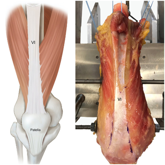
In all specimens, 3 distinct layers formed the QT. The first (superficial) layer was formed by the rectus femoris, which was fused to the second layer with an unclearly defined direct attachment to the patella. The median length of the QT was 86.9 mm (range, 68.4-98.9 mm). The second (middle) layer consisted of the vastus medialis and vastus lateralis and was found to have fibers running in an oblique direction that attached on the patella. A "fuse point," where the proximal part of the rectus femoris started to merge to the second layer, was identified at a median of 48.7 mm (range, 27.9-62.6 mm) from the PEL. The third (deep) layer consisted of the vastus intermedius. The median thickness of the graft centrally at 20, 40, 60, 80, and 100 mm from the PEL was 8.5, 7.2, 7.5, 6.5, and 5.4 mm, respectively.
Overall, 3 different layers of the QT were consistently found in all specimens. The first layer was fused with the second layer, and the direction of the fibers of the second layer or the vastus medialis and vastus lateralis was oblique. The median length of the QT was 86.9 mm, and the thickness of the tendon diminished proximally.
This study allows for a better understanding of QT anatomy when harvesting the tendon as a graft for ligamentous reconstruction.
由于股四头肌肌腱(QT)在重建前交叉韧带时作为移植物的使用增加,对其解剖结构的详细了解具有临床意义。
定性和定量描述年轻成人标本中QT的解剖结构。
描述性实验室研究。
本研究共使用了18个不成对的尸体膝关节,平均年龄为30.1岁(范围18 - 38岁)。使用三维坐标测量系统评估QT不同层之间的结构关系及其与髌骨的附着情况,并从髌骨关节面线(PEL;定义为髌骨内外侧关节面之间的直线)和近端以2厘米间隔在内侧、中央和外侧测量QT厚度。
在所有标本中,QT由3个不同的层组成。第一层(浅层)由股直肌形成,它与第二层融合,与髌骨的直接附着不明确。QT的中位长度为86.9毫米(范围68.4 - 98.9毫米)。第二层(中层)由股内侧肌和股外侧肌组成,发现其纤维呈斜向走行并附着于髌骨。在距PEL中位数为48.7毫米(范围27.9 - 62.6毫米)处确定了一个“融合点”,即股直肌近端开始与第二层融合的点。第三层(深层)由股中间肌组成。在距PEL 20、40、60、80和100毫米处中央的移植物中位厚度分别为8.5、7.2、7.5、6.5和5.4毫米。
总体而言,在所有标本中均一致发现QT有3个不同的层。第一层与第二层融合,第二层即股内侧肌和股外侧肌的纤维方向是斜向的。QT的中位长度为86.9毫米,肌腱厚度向近端逐渐减小。
本研究有助于在将肌腱作为韧带重建移植物取材时更好地理解QT的解剖结构。