Surgical Research LaboratoryDepartment of SurgeryUniversity Medical Center GroningenGroningenthe Netherlands.
Department of RadiologyUniversity Medical Center GroningenGroningenthe Netherlands.
Hepatology. 2022 Apr;75(4):898-911. doi: 10.1002/hep.32169. Epub 2021 Dec 5.
Portal vein thrombosis (PVT) is a common complication of cirrhosis. The exact pathophysiology remains largely unknown, and treatment with anticoagulants does not lead to recanalization of the portal vein in all patients. A better insight into the structure and composition of portal vein thrombi may assist in developing strategies for the prevention and treatment of PVT.
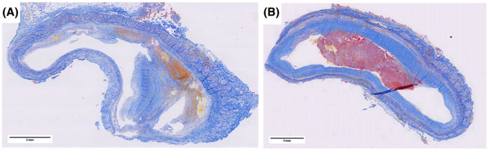
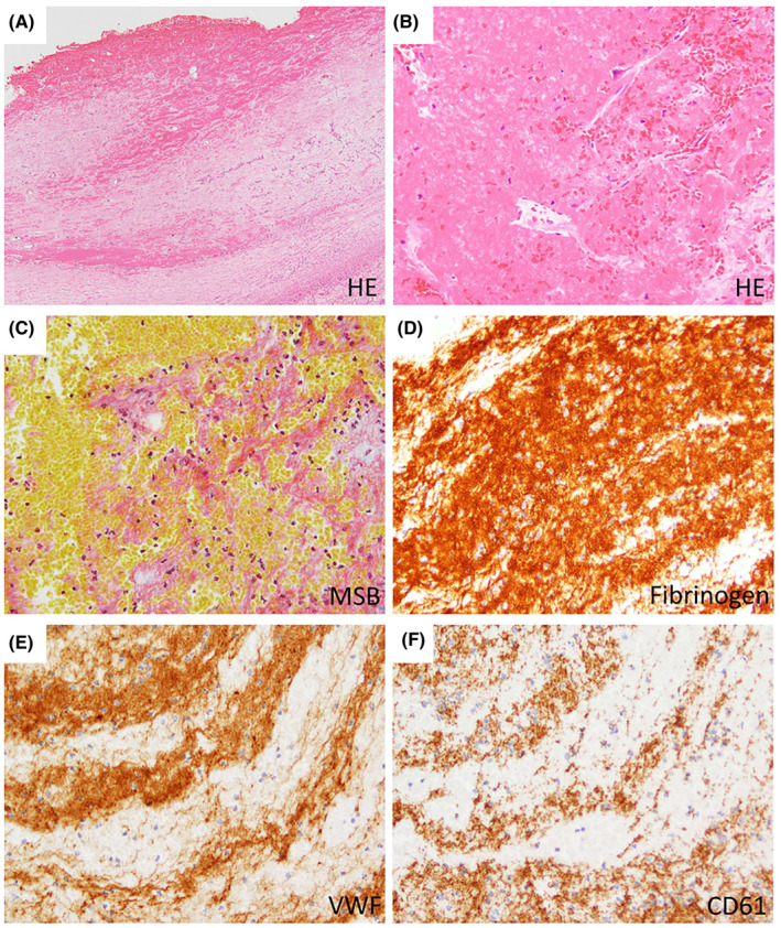
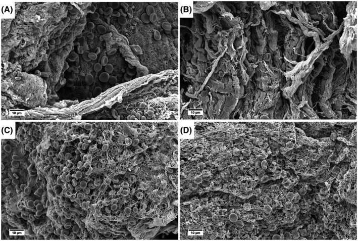
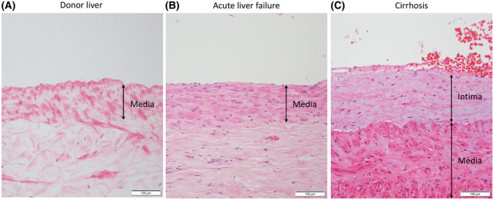
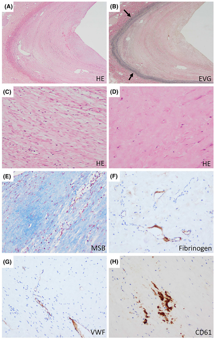
Sixteen prospectively and 63 retrospectively collected nonmalignant portal vein thrombi from patients with cirrhosis who underwent liver transplantation were included. Histology, immunohistochemistry, and scanning electron microscopy were used to assess structure and composition of the thrombi. Most recent CT scans were reanalyzed for thrombus characteristics. Clinical characteristics were related to histological and radiological findings. All samples showed a thickened, fibrotic tunica intima. Fibrin-rich thrombi were present on top of the fibrotic intima in 9/16 prospective cases and in 21/63 retrospective cases. A minority of the fibrotic areas stained focally positive for fibrin/fibrinogen (16% of cases), von Willebrand factor (VWF; 10%), and CD61 (platelets, 21%), while most of the fibrin-rich areas stained positive for those markers (fibrin/fibrinogen, 100%; VWF, 77%; CD61, 100%). No associations were found between clinical characteristics including estimated thrombus age and use of anticoagulants and presence of fibrin-rich thrombi.
We demonstrate that PVT in patients with cirrhosis consists of intimal fibrosis with an additional fibrin-rich thrombus in only one-third of cases. We hypothesize that our observations may explain why not all portal vein thrombi in patients with cirrhosis recanalize by anticoagulant therapy.
门静脉血栓形成(PVT)是肝硬化的常见并发症。其确切的病理生理学机制在很大程度上仍不清楚,且抗凝治疗并不能使所有患者的门静脉再通。对门静脉血栓的结构和成分有更深入的了解,可能有助于制定预防和治疗 PVT 的策略。
本研究前瞻性纳入了 16 例和回顾性纳入了 63 例因肝硬化行肝移植的非恶性门静脉血栓患者,使用组织学、免疫组织化学和扫描电子显微镜来评估血栓的结构和成分。对最近的 CT 扫描进行重新分析以评估血栓特征。将临床特征与组织学和影像学发现相关联。所有样本均显示出增厚的纤维化内皮层。在 9/16 例前瞻性病例和 21/63 例回顾性病例中,纤维蛋白丰富的血栓位于纤维化内皮层的顶部。少数纤维化区域局灶性地呈纤维蛋白/纤维蛋白原(16%的病例)、血管性血友病因子(VWF;10%)和 CD61(血小板,21%)阳性,而大多数纤维蛋白丰富的区域这些标志物均呈阳性(纤维蛋白/纤维蛋白原,100%;VWF,77%;CD61,100%)。未发现临床特征(包括估计的血栓年龄和抗凝治疗的使用)与纤维蛋白丰富的血栓之间存在关联。
我们证实,肝硬化患者的 PVT 由内皮层纤维化组成,仅三分之一的病例存在额外的纤维蛋白丰富的血栓。我们假设,我们的观察结果可能解释了为什么不是所有肝硬化患者的门静脉血栓通过抗凝治疗再通。